See how advanced slide-based imaging connects tissue architecture with cellular detail across fluorescent and histological samples.


Measuring mRNA and protein together at single cell resolution can uncover tumor-specific signaling activity and immune features.


Explore how integrating spatial analysis with flow cytometry can uncover new dimensions of CAR T cell biology.

Application Note
Complementary liquid chromatography techniques can improve the retention and separation of challenging radioligand therapy compounds.

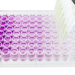
Discover practical strategies for performing reliable serial dilutions with optimized liquid handling and mixing.

Learn best practices for improving the accuracy, precision, and reproducibility of automated serial dilution workflows.
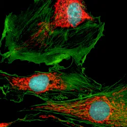
Understand how fluorophore performance and high-content imaging work together to support sensitive, reproducible multiplex imaging.

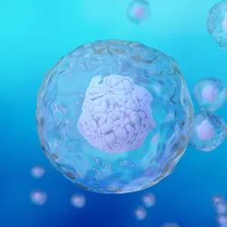
Advances in automated imaging are expanding the scalability of organoid phenotyping workflows.
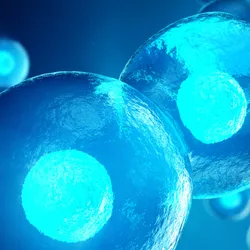
Imaging-enabled flow cytometry helps distinguish biologically relevant cell-cell interactions from coincident events to support immunotherapy research.
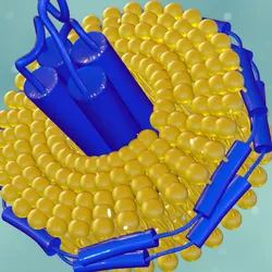
Mass photometry supports membrane protein characterization by providing rapid insights into sample composition, purity, and molecular assembly.

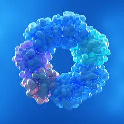
Automated mass photometry helps reveal the complex dynamics of protein oligomerization and the factors that govern protein assembly.
Explore how confocal live-cell imaging can reveal immune-tumor interactions and cytotoxic responses in three-dimensional tumor spheroid models.