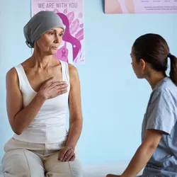
Cancer

| 3 min read
Patient advocates warn that political momentum to reduce animal testing is outpacing the scientific validation of alternative methods.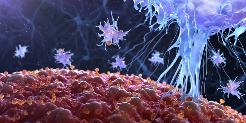
| 5 min read
Therapeutic cancer vaccines are being reengineered to deliver stronger immune activation and longer-lasting protection against disease recurrence.
| 5 min read
New population-level data reveal unprecedented declines in cervical cancer among vaccinated cohorts, even as vaccination rates begin to slip in some countries.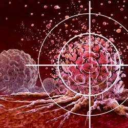
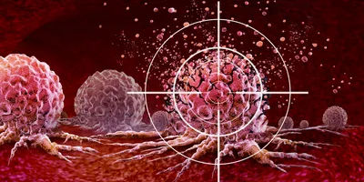
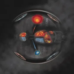
Researchers are uncovering new ways to attack some of cancer’s most elusive drivers.
| 6 min read
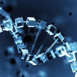
By stabilizing G-quadruplex DNA, a revived anticancer drug offers a new way to shut down a key oncogene.
| 4 min read
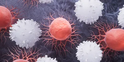
A rare immune cell subset with the capacity to self-renew, persist long-term, and drive potent antitumor responses
| 4 min read
Pharma deals, FDA review hurdles, and more led the news this week.
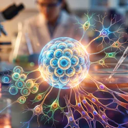
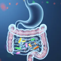
Gain insight into how microbiome research is advancing the understanding of cancer biology and therapeutic outcomes.
| 3 min read
Scientists at Johns Hopkins used a whole-body imaging technique to track where therapeutic cells go after injection

| 3 min read
Trained on more than 30,000 tumor genomes, MutationProjector offers drug developers a broader framework for connecting cancer genetics to therapy outcomes.
| 3 min read
As the field moves from fecal transplants toward defined bacterial consortia, the technical gaps in design, analytics, and co-cultivation are coming into sharper focus.

| 5 min read
Researchers identify a slow-cycling, therapy-tolerant state in breast cancer cells linked to Rac1 signaling and late metastatic relapse.