Explore how breakthroughs in fast confocal imaging and 3D cell culture converged to transform the study of complex human biology in vitro.

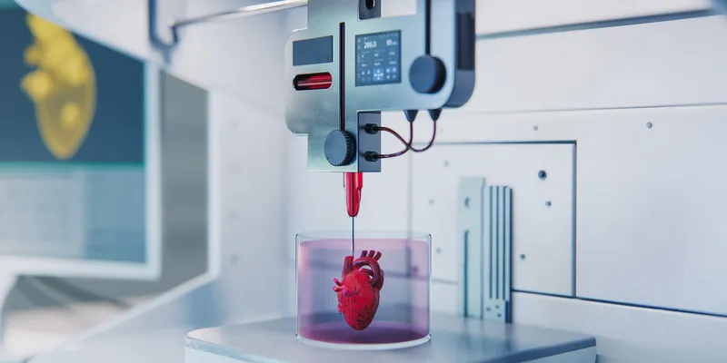
Discover the innovations that turned 3D fabrication into tools for building complex, lifelike biological structures.

| 1 min read
The milestone article will trace the controversial discovery of prostate-specific antigen (PSA) and the subsequent purification, characterization, and clinical use of PSA as a biomarker blood test for prostate cancer.Milestones
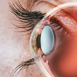
Over the past five decades, scientists have figured out ways to control the disease-fueling growth of blood vessels in the body.

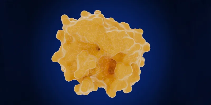
Researchers continuously push the boundaries of what’s possible with protein analysis tools.

| 1 min read
In a journey fraught with heartache and persistence, medical research giants paved the way for successful bone marrow transplantation.

| 1 min read
Since their discovery in the 1890s, viruses have intrigued scientists as potential cancer-killing agents.

Interdisciplinary collaboration and innovation propelled transformative advancements in single cell proteomic analysis.

Gene therapy has a complicated history, marked by many ups and downs and crafted by ever-advancing technologies.

| 1 min read
In the 1930s, HPV
wasn’t on the radar for most scientists.Four decades passed before anyone connected
HPV with cervical, penile, or anal cancer, and it took even longer before scientists
developed a prophylactic vaccine.

| 1 min read
The discovery of insulin and its role in diabetes began with extracting canine pancreatic secretions and culminated in the first artificial pancreas in 2016.

| 1 min read
Transgenic mice play pivotal roles in scientific and therapeutic discovery, but how did they become a staple model system?