Multimedia & Resources

A researcher wearing a white lab coat and blue nitrile glove holds a tiny brown glass sample vial with a blue cap in front of an autosampler carousel on a HPLC laboratory analyzer.
Hear an expert perspective on the hidden variables affecting chromatographic methods and how to build more robust, reliable workflows.
3D rendering of a Y-shaped antibody molecule surrounded by blurred molecular structures against a red background.
Explore how charge variant fractionation combined with downstream analysis methods can reveal molecular differences in complex biotherapeutics.
A 3D medical illustration of a cancer cell or tumor, featuring a main red mass covered in bumpy white textures with long, root-like tentacles branching outward across a gray surface.
Available on DemandLearn how patient-derived tumor organoids, AI, and phenotypic screening are advancing the discovery of small molecule therapeutics.
A scientist in a white lab coat looking into a microscope in a brightly lit modern laboratory.
Available on DemandLearn how developmental and reproductive toxicology study selection supports regulatory decision-making and generates meaningful nonclinical safety data.
Researcher using a laptop with a digital DNA helix and molecular biology graphics overlaid, illustrating connected workflows for sequence design, data management, and therapeutic research.
To keep pace with modern drug discovery, researchers need molecular biology approaches that can support complexity without slowing down the science.
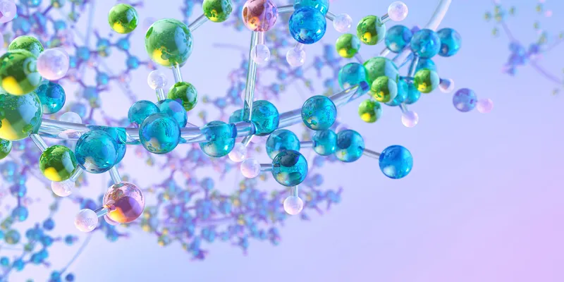
A colorful 3D rendering of molecular structures featuring interconnected blue, green, and yellow spheres representing atoms and bonds, symbolizing chemical and biological complexity.
In parallel with advances in new modalities and digital design, synthetic chemistry remains integral to pharmaceutical innovation.
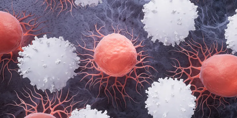
An illustration showing red cancer cells surrounded by white immune cells interacting on a dark textured background.
A powerful tool in modern oncology, immunotherapy doesn’t work the same for everyone. Researchers are exploring why and developing ways to improve its effectiveness.
Antibodies are shown as light pink and orange Y-shaped molecular structures against a light blue and purple background.
Monoclonal antibodies are life-saving therapeutics that can be used for a myriad of conditions from infectious diseases to cancer.  
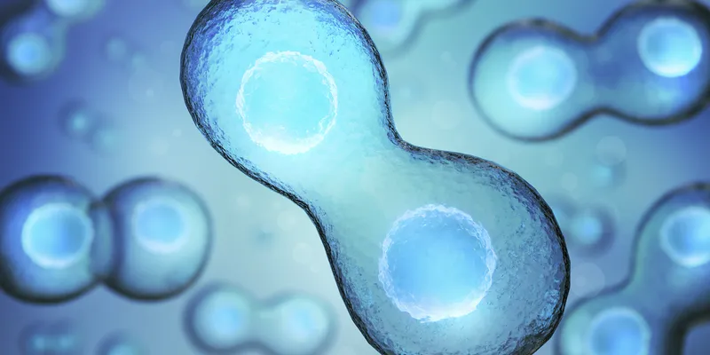
Illustration of a cell undergoing division, showing two connected daughter cells with visible nuclei, rendered in blue tones
Explore how breakthroughs in fast confocal imaging and 3D cell culture converged to transform the study of complex human biology in vitro.
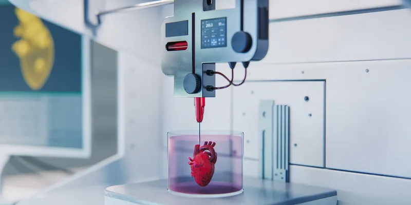
A bioprinting nozzle deposits material into a transparent container holding a heart-shaped tissue construct in a laboratory setting.
Discover the innovations that turned 3D fabrication into tools for building complex, lifelike biological structures.
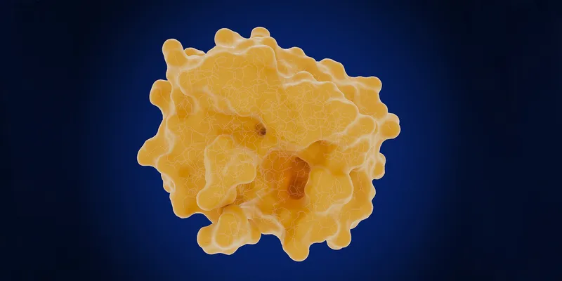
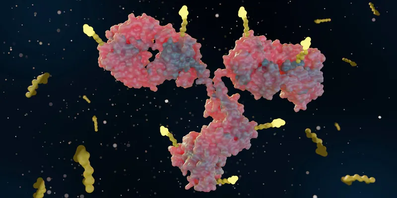
A 3D molecular rendering of the prostate-specific antigen, showing its folded yellow protein structure with a large cavity representing the active site on the lower right.
| 1 min read
The milestone article will trace the controversial discovery of prostate-specific antigen (PSA) and the subsequent purification, characterization, and clinical use of PSA as a biomarker blood test for prostate cancer.
 Close-up of a human eye with a medical illustration overlay showing the internal anatomy, including the cornea, retina, and optic nerve.
Over the past five decades, scientists have figured out ways to control the disease-fueling growth of blood vessels in the body.
A toddler sits on a woman’s lap. Both look at a smart phone propped up on the table in front of them.
| 9 min read
A new app relies on smartphone cameras and algorithms to check for signs of neurodivergence in children.
A microscopic view of the human heart muscle tissue.
| 10+ min read
After a heart attack, the damage is permanent — for now. Researchers are testing innovative ways to get the heart to regenerate muscle cells and fix itself.
A photo of a 17th century herbal medicine book from The Huntington Library’s collection.
| 10+ min read
Could the solution to antibiotic resistance already have been discovered? Microbiologists and historians probe medieval and ancient texts to find out.
A series of four eye irises are arranged in a line against a black background.
| 8 min read
In diabetic macular edema, senescent cells build up in the retina, leading to vision loss. A new drug forces these undead cells to die, healing the retina.
Stylized three-dimensional rendering of multicellular spheroids enclosed within transparent structures.
Navigate the transition from two-dimensional to phenotypic three-dimensional cell models for drug discovery.
Gloved researcher transferring liquid into a microplate using a multichannel pipette.
Discover practical strategies to improve pipetting accuracy, reproducibility, ergonomics, and instrument performance across diverse laboratory workflows.
A 3D rendering illustrates a sandwich ELISA technique, where antigen detection is achieved between two layers of antibodies: a capture antibody and a detection antibody
Learn the key characteristics that determine whether an immunoassay generates accurate and reproducible data.
A conceptual scientific illustration featuring a close-up of a dark metallic pocket watch or stopwatch face to symbolize time-lapse analysis.
Explore how real-time live-cell analysis supports cell culture quality, 3D model characterization, and data-driven biological insights.
Digital illustration of a magnifying glass enlarging a red tumor composed of clustered cells.
Explore how patient-derived 3D models and multiomics analyses can help investigate drug response, resistance mechanisms, and precision oncology approaches.
Shaping Science graphic featuring the question “How can labs become truly sustainable?” and a photo of James Connelly, Chief Executive Officer of My Green Lab.
| 1 min read
Creating more sustainable laboratories depends on practical changes that strengthen scientific performance while reducing environmental impact.
Image of DNA being edited.
| 1 min read
Philip Wills, Chief Commercial Officer of the Cell & Gene Therapy Division at Catalent, reflects on how the gene therapy industry is evolving from early scientific breakthroughs to commercial maturity.
Why sample quality can make or break structural biology
| 1 min read
Discover why mass photometry is becoming essential in the toolbox of protein studies.
Illustration of interconnected blue molecular structures with spherical atoms and chemical bonds on a light blue background.
Discover how analyte properties can guide column selection and help determine the most effective chromatographic approach.
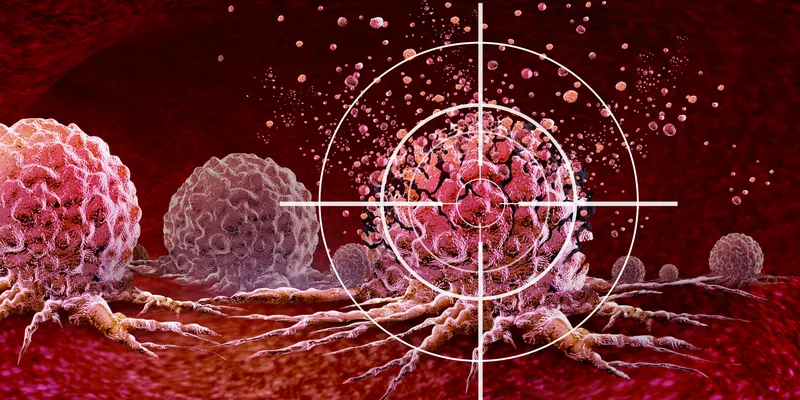
Ultrasound Cancer Treatment and new Cancer killing tumours therapy with vibrating molecules breaking the membranes of malignant tumor cells as an oncology therapy fighting Cancerous Growth and Metastasis.
Researchers are uncovering new ways to attack some of cancer’s most elusive drivers.
Automated liquid handling tips dispense samples into a multiwell microplate during a high-throughput screening experiment, with colorful fluorescent assay signals blurred in the background.
Learn the key assay design principles and workflow considerations that help support robust, scalable, and reliable high-throughput screening campaigns.
3D illustration, abstract pathogen as a type of flu - H1N1, hepatitis viruses, influenza virus, flu, aids. Virus abstract background. Virus infects human cells. Infection causing chronic disease
Learn how upstream analytical strategies can improve AAV yield, quality, and process efficiency.
A detailed image generated by SILA-integrated microscopy of the diverse biological structures of a lily flower
| 1 min read
A collaborative team overcomes the long-standing challenge of blurred imaging, illuminating once hidden details within the depths of thick samples.

Subscribe to Newsletter

Subscribe to our eNewsletters

Stay connected with all of the latest from Drug Discovery News.

Subscribe

Sponsored

Colorful geometric graphic panel featuring white line illustrations of the human heart, brain, kidneys, pancreas, lungs, stomach, uterus, colon, and bladder, arranged in a grid of red, blue, and purple shapes.
Learn how biologically relevant 3D human tissue models can reveal organ-specific toxicity earlier and support more informed safety assessment.
A 3D molecular model showing interconnected red, blue, and silver atoms against a gray background.
Explore chromatographic strategies for retaining, separating, and analyzing challenging polar compounds across pharmaceutical applications.
A conceptual illustration of molecular structures and chemical formulas representing metabolites and their analysis.
Explore the science of metabolomics and drug metabolites, their roles as biomarkers, and the analytical methods for their characterization.