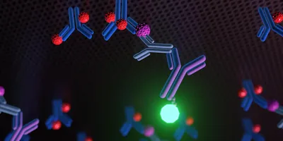
Learn the key characteristics that determine whether an immunoassay generates accurate and reproducible data.


Explore how real-time live-cell analysis supports cell culture quality, 3D model characterization, and data-driven biological insights.

Explore key considerations for improving molecular biology workflows through centralized design, data management, and collaborative research practices.

Ebook
Advanced mass spectrometry and multidimensional separation strategies are improving the characterization of proteins, peptides, and complex biotherapeutics.


| 1 min read
The toughest problem to solve in drug development may not be finding new medicines, but building the infrastructure to make them reliably.
Discover how adaptive PCR workflows can improve sequencing consistency and data quality across fragmented, low-input, and variable-quality samples.

Discover workflow and fragmentation techniques that enhance metabolite identification and support more reliable structural interpretation.

From study design to biomarker interpretation, discover what it takes to incorporate metabolomics effectively into drug discovery research.

Advances in high-dimensional flow cytometry and integrated imaging are enabling deeper, more reproducible insights into cellular behavior.

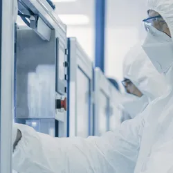
Explore key strategies for achieving reliable, compliant sterility testing in pharmaceutical labs.
| 1 min read
Our March 2026 issue explores the ideas, challenges, and innovations influencing the future of drug discovery.
Explore how kinetic measurements provide deeper insight into drug–target interactions beyond affinity alone.