Yuning Wang, PhD
Yuning joined the custom content team at Drug Discovery News in June 2022. She earned her PhD in biochemistry from the University of Western Ontario, where she investigated how calcium sensor proteins regulate muscle cell membrane repair and cause muscular dystrophy. Yuning developed a passion for science communication during graduate school and began her career as a science writer in 2020. She enjoys reading, gardening, and trying new restaurants in Toronto.
Articles by Yuning Wang, PhD
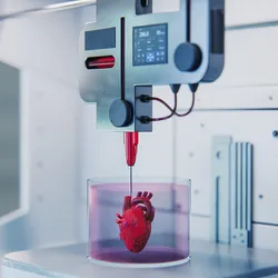
Discover the innovations that turned 3D fabrication into tools for building complex, lifelike biological structures.

| 1 min read
As drug discovery grows more complex, the pace of discovery increasingly depends on the tools and people working behind the scenes.
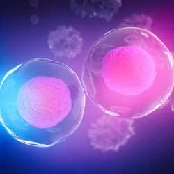
Explore evolving technologies, analytical strategies, and expert guidance supporting high-quality flow cytometry research.
| 1 min read
The milestone article will trace the controversial discovery of prostate-specific antigen (PSA) and the subsequent purification, characterization, and clinical use of PSA as a biomarker blood test for prostate cancer.
| 1 min read
Laboratories account for a surprising share of global emissions and plastic waste, making sustainability a priority for modern research.
| 1 min read
Why is spatial proteomics in the spotlight? Learn how mapping proteins in place is changing the way researchers study cells, diseases, and potential new drugs.

| 2 min read
Autophagy in cancer is a double-edged sword: It protects cells from malignant transformation, but once tumors form, it helps them thrive.

| 1 min read
Born to differentiate and heal, mesenchymal stem cells carry great potential for treating a wide spectrum of diseases.

| 7 min read
Achieving higher accuracy and depth in bottom-up proteomics involves navigating a multifaceted process with various steps and challenges.

From signaling hunger to influencing mood, the vagus nerve keeps the gut and brain in sync.

| 1 min read
Since their discovery in the 1890s, viruses have intrigued scientists as potential cancer-killing agents.

| 10+ min read
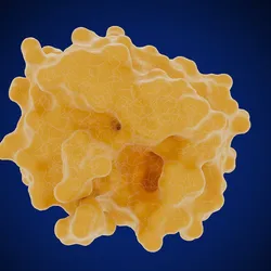
Learn how structure-based drug design uses a protein's 3D structure to create precisely targeted medicines. Explore the key techniques, from X-ray crystallography to AI-powered modeling.