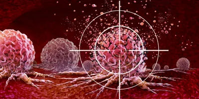
A tumor is much more than a lump of cancer cells. It houses immune cells, blood vessels, and an extracellular matrix, which are key to the disease’s development and progression. Yet, traditional flat cultures fail to capture the complexity of the tumor microenvironment and the interactions among its various components. While some 3D models such as organoids are better at mimicking this intricate ecosystem, they still lack some of these vital components and are not highly reproducible. Because of this, some scientists turned their attention to bioprinting technologies.
When I saw a bioprinter, I thought it was an interesting way of being able to make more tissues in a shorter period of time.
- Stephanie Willerth, University of Victoria
3D bioprinters operate much like traditional 3D printers, building solid objects by layering material in a predesigned pattern. The twist lies in the “bio” of bioprinting, where bioinks replace traditional inks. Instead of thermoplastic materials, bioinks consist of living cells, active molecules, or biomaterials such as alginate or collagen. Ideally, bioinks mimic the mechanical and biological properties of the target tissues, so to model tumors, researchers need to know the cell types that exist in the cancer microenvironment as well as its extracellular matrix components.
Replicating the heterogeneous architecture of solid cancerous tumors opens a world of possibilities for exploring tumor biology. For example, researchers can investigate the individual or combined roles of each cell type in tumor growth, aggressiveness, or metastatic properties. The approach may help scientists identify the components within this microenvironment that contribute to therapy resistance or that induce blood vessel formation within the tumor. Furthermore, bioprinted models could serve as platforms for screening drugs in a high-throughput manner, assessing their efficacy, and contributing to the development of precision medicine approaches.
Beyond flat and round cultures: the power of ink
While growing human cancer cell lines in a dish is a practical way to study cancer physiology, experiments in these oversimplified models may not always translate successfully to animal models, much less to clinical applications. The advantages of bioprinted models over flat cultures might seem evident, but what about other non-printed 3D models, such as organoids or spheroids?
For many years, University of Victoria’s biomedical engineer and bioprinting expert Stephanie Willerth has engineered 3D neural tissue from stem cells to study neurological disorders such as Alzheimer’s and Parkinson’s diseases. But before moving into bioprinting technologies, her team created these structures manually. For example, they used a pipet to encapsulate spheroids into 3D scaffolds. “When I saw a bioprinter, I thought it was an interesting way of being able to make more tissues in a shorter period of time,” she said. “I was also intrigued by the idea of actually placing certain types of cells on certain areas.”

Controlling the location of each cell subpopulation is one of the key advantages of bioprinted models compared to other 3D techniques. It allows for a more precise model architecture than that achieved in organoids, biomaterial scaffolds, or cancer-on-chip systems (1). In bioprinted models, “You can control cell-cell interactions by facilitating the right positions of specific cell types,” said İbrahim Özbolat, a bioengineer and bioprinting specialist at Pennsylvania State University. This predefined local arrangement also makes bioprinted 3D structures more homogeneous compared to organoids, he said. Willerth added, “[Bioprinted models] tend to be more reproducible than organoids.” \
Other 3D models also may miss some components of the tumor microenvironment (2). They might include fewer cell types or lack the extracellular matrix. Many of these models also fail to accurately mimic how blood vessels grow and infiltrate the tumor (3). In contrast, “You can induce vascularization with the 3D bioprinting,” Özbolat noted, which is another key advantage. While it is also possible to manually achieve vascularization in organoids, he acknowledged, the controlled printing approach allows extra capabilities. “You can build some channel structures where you can perfuse and make blood vessel-like organization.”
However, 3D bioprinted models also have limitations and fail to represent other tumor properties. The native tumor starts with the gradual growth of cancer stem cells, and throughout that process, it recruits other cell types from the neighborhood. “Everything has some spatial as well as some temporal interactions, versus in the printing process, [where] you have all the cell types and then you put all these together,” Özbolat said. The model may recapitulate the spatial arrangement, but not the temporal one. “You still miss that important aspect in your 3D printed model,” he noted.
Bioprinting versus animal testing
Animal models are another vital tool in cancer research, but they do not entirely represent human physiology. “One of the main ways people test anti-cancer therapeutics right now is to put a tumor in a mouse that lacks an immune system,” said Willerth. “But the immune system plays a huge role, whether it’s regulated or dysregulated, on how we respond to cancer. So, it’s not a very good system for testing these drugs.”
The use of animal models also raises ethical concerns (4). In this sense, some experts view bioprinted structures as a means to reduce and potentially replace animal experimentation (5). In addition to facilitating the inclusion of healthy human tissue, these structures reduce experimentation time and costs and offer greater control.
Özbolat emphasized that, for now, researchers still need to test most cancer therapies in animal models before moving into clinical translation. “There are a lot of different things contributing to cancer that exist in the live body versus in the 3D model,” he said. “We’re not really 100 percent sure about the 3D model, its capabilities, or its relevance to native biology, so that’s why we still have some more confidence if we test on the animal model as well.”
Printing tumors for refining cancer therapies
In the landscape of cancer drug development where the probability of success for drugs entering clinical trials is below ten percent, a model with the characteristics of bioprinted tumors is promising (6,7). For instance, high-throughput drug screening in these 3D models would enable a faster transition to the animal testing phase in addition to offering higher confidence for drug efficacy (8).
Since the properties of the extracellular matrix and other components of the tumor microenvironment influence drug diffusion, bioprinted tumors offer superior models for testing therapies compared to flat cell cultures. Based on several studies on printed tumors, researchers have already questioned findings from 2D experiments, particularly regarding treatment sensitivity and resistance. For example, many chemotherapy drugs such as cisplatin and 5-fluorouracil are highly effective at killing cultured cancer cells in a monolayer, but their efficacy reduces both in 3D models and in mice with patient-derived tumors (9,10).

Willerth and her colleagues have also explored how drugs behave using different modeling structures. Her team has a strong focus on neurodegenerative diseases, but they got into bioprinting cancer models when Chris Lee, who was a talented undergraduate student at the time, approached her to join the lab. “He really wanted to work on cancer, so we decided to work on brain cancer.”

The team developed a bioprinted glioblastoma tumor model (11). “We made a printable version of fibrin, which is the protein that clots your blood,” explained Willerth, who is also the cofounder and chief executive officer of the bioink developer company Axolotl Biosciences. “We got really huge, lovely tumors,” she added.
The team tested the glioblastoma cell responses to a small molecule cocktail that aims to reprogram cancer cells into nonproliferating neurons. While the treatment impaired the cells’ proliferation abilities in the 3D tumor, the cancer cells showed higher resistance to the treatment compared to those grown in a dish (12). “It’s super easy to kill cancer in 2D; it’s much harder to kill it when it’s a tumor,” Willerth said.
Her team plans to continue comparing cell responses in such different settings for other drug treatments (13). “Those comparisons are really important to make for [3D bioprinted] models really to be adoptable,” she argued.
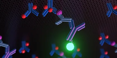
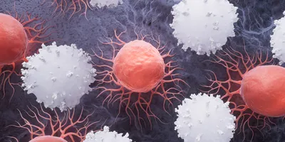
Bioprinted models may also aid in understanding why some therapies are ineffective against certain cancers and in devising ways to improve them. CAR T cell therapies, for example, are great at fighting blood cancers, but they are not as effective against solid tumors. In blood cancers, doctors inject the T cells directly into the patient, and the engineered cells circulate throughout the body to meet, interact with, and kill the cancer cells. For solid tumors, T cells need to find the cancer cells and then infiltrate into a very dense environment. Özbolat compared this search to a person looking for a gas station in different surroundings. “You can always find a gas station next to the highway easier [than] in the middle of downtown,” he said.
There are a lot of different things contributing to cancer that exist in the live body versus in the 3D model.
- İbrahim Özbolat, Pennsylvania State University
Özbolat and his colleagues were interested in modeling the crowded environment CAR T cells face when treating solid tumors. They bioprinted a breast tumor that included various representative cell types, such as vein endothelial cells and vasculature (14). They then perfused the 3D model with CAR T cells. They varied the number of CAR T cells they delivered, treatment duration, and target antigens. By modeling these processes, they learned how the CAR T cells attach to the innermost layer of the blood vessels and then migrate to finally invade the tumor and kill it. Since infiltration is one of the main limitations of CAR T cell therapies for treating solid tumors, a better understanding of this process in different treatment conditions may help improve these therapies.
The next question is whether scientists can develop more personalized therapies based on these 3D models, Özbolat said, for example, by printing tumors derived from patient-specific cells. This approach could help predict the dosage and the drug cocktail for each person or identify a cancer’s susceptibility or resistance to a given therapy (15). Tumor bioprinting, therefore, holds the promise to potentially guide clinical decisions towards optimal cancer treatments in a timely and more efficient manner than current practices.
References
- Sharma, R. et al. 3D bioprinting complex models of cancer. Biomater Sci 11, 3414-3430 (2023).
- Neufeld, L. et al. 3D bioprinted cancer models: from basic biology to drug development. Nat Rev Cancer 22, 679-692 (2022).
- Germain, N. et al. Current Advances in 3D Bioprinting for Cancer Modeling and Personalized Medicine. Int J Mol Sci 23, 3432 (2022).
- Ferdowsian, H.R. & Beck, N. Ethical and Scientific Considerations Regarding Animal Testing and Research. PLoS One 6, e24059 (2011).
- Sztankovics, D. et al. 3D bioprinting and the revolution in experimental cancer model systems—A review of developing new models and experiences with in vitro 3D bioprinted breast cancer tissue-mimetic structures. Pathol Oncol Res 29, 1610996 (2023).
- Hay, M. et al. Clinical development success rates for investigational drugs. Nat Biotechnol 32, 40-51 (2014).
- Wong, C.H. et al. Estimation of clinical trial success rates and related parameters. Biostatistics 20, 273-286 (2019).
- Mazzocchi, A. et al. 3D bioprinting for high-throughput screening: Drug screening, disease modeling, and precision medicine applications. Appl Phys Rev 6, 011302 (2019).
- Abreu Miranda, M. et al. Cytotoxic and chemosensitizing effects of glycoalkaloidic extract on 2D and 3D models using RT4 and patient derived xenografts bladder cancer cells. Mater Sci Eng C Mater Biol Appl 119, 111460 (2020).
- Mao, S. et al. Bioprinting of patient-derived in vitro intrahepatic cholangiocarcinoma tumor model: establishment, evaluation and anti-cancer drug testing. Biofabrication 12, 045014 (2020).
- Lee, C. et al. Bioprinting a novel glioblastoma tumor model using a fibrin-based bioink for drug screening. Mater Today Chem 12, 78-84 (2019).
- Lee, C. et al. Direct Reprogramming of Glioblastoma Cells into Neurons Using Small Molecules. ACS Chem Neurosci 9, 3175-3185 (2018).
- Smits, I.P.M. et al. Novel N-cadherin antagonist causes glioblastoma cell death in a 3D bioprinted co-culture model. Biochem Biophys Res Commun 529, 162-168 (2020).
- Dey, M. et al. Chemotherapeutics and CAR-T Cell-Based Immunotherapeutics Screening on a 3D Bioprinted Vascularized Breast Tumor Model. Adv Funct Mater 32, 2203966 (2022).
- Yi, H.-G. et al. A bioprinted human-glioblastoma-on-a-chip for the identification of patient-specific responses to chemoradiotherapy. Nat Biomed Eng 3, 509-519 (2019).