Articles, Explainers, Milestones
| 5 min read
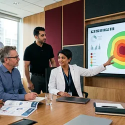
Precision oncology has focused on mutations. Spatial profiling adds the microenvironmental context that increasingly explains why matched therapies still fail.
| 5 min read
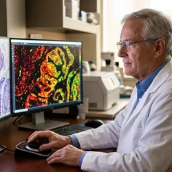
Spatial biology and digital pathology are converging on the same tissue slide. Their merger is what will actually bring spatial insights to clinical decisions.
| 5 min read
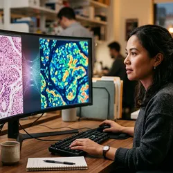
Pathology has always been spatial. Now it is becoming molecular and computational too, and the fusion of the two is a genuinely new diagnostic substrate.
| 5 min read
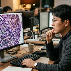
The scale of spatial pathology data makes AI not optional but necessary. Deep learning is how tissue maps become clinical predictions.
| 5 min read
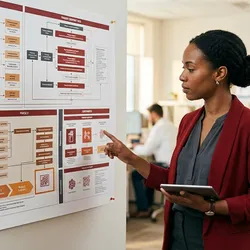
Spatial assays are moving into trial protocols, first as exploratory endpoints, increasingly as the basis for who gets enrolled and why.
| 4 min read
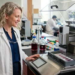
Spatial biology’s move from boutique experiment to pipeline tool depends on automation. This is how pharma is industrializing it.

| 5 min read
Spatial assays are complex, multi-step, and image-dependent, exactly the profile regulators scrutinize hardest. Knowing the path early shapes development
| 5 min read
A spatial biomarker is not just what is present in a tissue. It is what is present, where, and next to what. That extra dimension is where the predictive power lives.
| 5 min read
Immuno-oncology’s next generation of targets and biomarkers is emerging from spatial data, where the interactions checkpoint drugs act on are finally visible.
| 6 min read
The next generation of biomarkers will not be single molecules but spatial patterns. Getting from a promising pattern to an approved diagnostic is its own discipline.
| 5 min read
PD-L1 status is a blunt predictor. Spatial context, which cells express it and next to what, explains far more about who actually responds.
| 5 min read
New imaging analysis from the Phase 2a trial of a new small molecule drug shows dose-dependent preservation of whole-brain connectivity.