- Key Takeaways
- A class B receptor built for a peptide
- How does GLP-1R send its signal?
- Biased agonism: one receptor, different signals
- Where is the GLP-1 receptor actually found?
- The brain: appetite, satiety, and reward
- How does GLP-1R act on the heart and kidney?
- The gut and beyond: GI effects and other tissues
- Why does receptor biology matter for drug discovery?
- Why this matters for drug discovery
GLP-1 receptor biology explains why a single class of drugs can lower blood sugar, curb appetite, and protect the heart and kidneys at once. The receptor is a class B G-protein-coupled receptor that signals mainly through cyclic AMP, and it is expressed far beyond the pancreas, in the brain, gut, vasculature, heart, and kidney. Understanding that structure, signaling, and distribution is the foundation for understanding the drugs themselves.
Key Takeaways |
|
A class B receptor built for a peptide
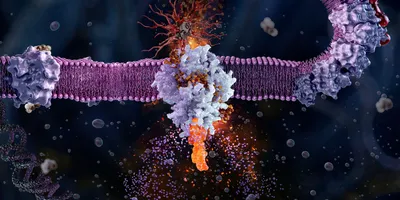
The GLP-1 receptor belongs to the class B1, or secretin, family of G-protein-coupled receptors, a group built to recognize peptide hormones rather than small molecules. Its architecture reflects that job: a large extracellular domain that captures one end of the peptide, joined to the seven-transmembrane core shared by all GPCRs. The peptide binds by a two-domain mechanism, with its C-terminus held by the extracellular domain while its N-terminus inserts into the transmembrane bundle to switch the receptor on.
That activation step was resolved in detail when researchers reported the first near-atomic cryo-electron microscopy structure of the activated, full-length GLP-1 receptor bound to its Gs protein. The structure showed the peptide clasped between the two domains and a sharp kink forming in the middle of transmembrane helix 6, which swings the receptor’s intracellular half outward to make room for the G protein. This is the molecular event that begins every GLP-1 signal.
This biology sits at the foundation of the wider story of GLP-1 and metabolic disease, which connects the receptor to the medicines built on it.
How does GLP-1R send its signal?
Once the receptor opens to the G protein, it couples mainly to the stimulatory G protein Gs, which activates adenylyl cyclase and raises intracellular cyclic AMP. That rise in cyclic AMP is the central second messenger of GLP-1 action, working through protein kinase A and the exchange protein Epac2 to drive the receptor’s downstream effects.
In the pancreatic beta cell, that cyclic AMP signal is what links GLP-1 to insulin: it amplifies glucose-stimulated insulin secretion without triggering it on its own, which is why the effect is glucose-dependent and carries little risk of hypoglycemia. The same receptor can also mobilize intracellular calcium and activate the ERK pathway, giving GLP-1 a signaling repertoire broader than cyclic AMP alone.
Biased agonism: one receptor, different signals
Signaling does not stop at the G protein. The activated receptor also recruits beta-arrestins, which dampen G protein signaling and draw the receptor inside the cell, a process that controls how long and how strongly the receptor fires. Different agonists tip this balance differently, favoring G protein signaling or arrestin recruitment to varying degrees, a phenomenon called biased agonism.
The balance matters because it shapes the biological response. Evidence suggests that favoring sustained G protein signaling over arrestin-driven internalization can prolong cyclic AMP production and insulin secretion, which has made biased agonism a deliberate goal in molecular design rather than an academic curiosity.
Turning that mechanistic insight into medicines is the province of applied discovery, traced in how this receptor biology was turned into a drug-discovery platform. The point here is simply that the receptor offers more than one signaling lever to pull.
Map where the receptor sits, and you have mapped where the drugs act.
Where is the GLP-1 receptor actually found?
For years, this was one of the hardest questions in the field, because receptor localization was confounded by nonspecific antibodies that produced false signals in tissues that turned out not to express the receptor at all. The picture only became reliable once researchers applied antibodies validated against knockout tissue, RNA in situ hybridization, and transgenic reporter models.
Using an extensively validated monoclonal antibody, one landmark study mapped the receptor to beta cells in the pancreas, the myocytes of the sinoatrial node in the heart, and the smooth muscle of arteries and arterioles in the kidney and lung. The table below summarizes where the receptor sits and what it does in each location.
Table 1. GLP-1 receptor distribution and function by tissue
Tissue | Where the receptor sits | What it does there |
Pancreas | Beta cells (strong), acinar cells (weak), not ducts | Glucose-dependent insulin release |
Brain | Arcuate and paraventricular nuclei, area postrema, brainstem | Appetite suppression, satiety, and nausea signaling |
Stomach and duodenum | Brunner’s glands, parietal cells, and gastric smooth muscle | Slowed gastric emptying, secretion control |
Heart | Sinoatrial node and atrial myocytes | Heart-rate increase, atrial natriuretic peptide release |
Kidney and vasculature | Smooth muscle of arteries and arterioles | Vascular tone, blood pressure, renal hemodynamics |
Immune system | Macrophages, lymphocytes, iNKT cells | Anti-inflammatory signaling |
The brain: appetite, satiety, and reward
The central nervous system is where much of the modern interest in GLP-1 lives, because the brain is where appetite is controlled. Validated mapping places the receptor in the arcuate and paraventricular nuclei of the hypothalamus, the area postrema, and the nucleus of the solitary tract in the brainstem, regions that govern hunger, satiety, and nausea. Activation there reduces food intake, and it is this central action, as much as any peripheral effect, that drives the weight loss seen with GLP-1 drugs.
The same circuitry explains a common side effect. The area postrema and brainstem nuclei that signal satiety also mediate nausea, which is why appetite suppression and gastrointestinal upset so often travel together. Receptor biology, in other words, predicts both the benefit and the burden of these drugs in the brain.
How does GLP-1R act on the heart and kidney?
The cardiovascular and renal benefits of GLP-1 drugs are real and now well documented in outcome trials, but the receptor biology behind them is more indirect than it first appears. In the heart, validated studies find the receptor concentrated in the sinoatrial node, which explains the modest increase in heart rate, and in atrial myocytes, where it stimulates the release of atrial natriuretic peptide. The working muscle of the ventricles shows little direct receptor expression.
The kidney tells a similar story. Rather than sitting on the filtering cells of the nephron, the receptor is found mainly in the smooth muscle of the renal vasculature, so much of GLP-1’s renal effect is hemodynamic and hormonal rather than a direct action on kidney tubules. The benefits are genuine, but they flow through the vasculature, the nervous system, and secondary mediators more than through the organ’s working cells, a distinction that matters when designing drugs meant to target these tissues.
The gut and beyond: GI effects and other tissues
In the gastrointestinal tract, the receptor appears in the Brunner’s glands of the duodenum, in parietal cells, and in gastric smooth muscle, where it slows gastric emptying. That single action prolongs the sense of fullness, but it is also the direct source of the characteristic gastrointestinal side effects of the GLP-1 class, from nausea to constipation. The same biology delivers the benefit and the tolerability problem.
Beyond these major organs, the receptor turns up on cells of the immune system, including macrophages, lymphocytes, and invariant natural killer T cells, where it appears to exert anti-inflammatory effects. This wider distribution is part of why GLP-1 drugs keep finding new candidate indications, though each new tissue claim now demands the same rigorous validation that corrected the earlier record.
Why does receptor biology matter for drug discovery?
Receptor biology is not background to the GLP-1 story; it is the explanation for almost every feature of the drugs. The glucose-dependence that makes them safe, the central action that drives weight loss, the cardiovascular and renal benefits, and the gastrointestinal side effects all trace back to where the receptor sits and how it signals. A discovery program that understands the receptor can anticipate effects rather than discover them in the clinic.
It also points to the frontier. Biased agonism offers a way to tune the signal, the multi-organ distribution suggests indications still being explored, and the lesson of the antibody era, that a receptor claim is only as good as its validation, applies to every new tissue and target. The biology, in short, is where the next generation of these drugs will be designed.
Why this matters for drug discovery |
If you are evaluating a GLP-1 program, start with the receptor. Its two-domain peptide binding constrains what a molecule must do to activate it, its Gs and arrestin signaling define the levers available through biased agonism, and its validated tissue distribution predicts both the therapeutic reach and the side-effect profile. Treat new tissue or indication claims with the skepticism the field learned the hard way. A receptor detected by an unvalidated antibody is a hypothesis, not a target, and the difference has already redrawn the map of where GLP-1 truly acts. |
This article was produced under Drug Discovery News’ AI Editorial Guidelines.