biochemistry

Available on Demand
Discover how new insights into a tardigrade-derived protein are informing new approaches to DNA protection and genome stability.
Available on Demand
Discover how rapid viscosity and rheology testing with minimal sample volumes can help make better formulation decisions.

| 2 min read
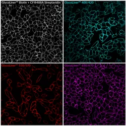
New GlycoLiner kits enable rapid, covalent labeling of live-cell surface glycoproteins using gentle, self-catalyzing chemistry at neutral pH.
| 3 min read
Researchers challenge decades-old assumptions about fragile protein cores, opening new paths for faster drug and enzyme design.

| 3 min read
In vivo studies of the antibiotic darobactin D22 against high-priority gram-negative bacteria show promise for further drug development.

| 3 min read
The new complex is inexpensive to produce, and its unique architecture makes chemical reactions integral to drug discovery more stable and efficient.

| 4 min read
A new wound dressing monitors infections in real time and administers timely treatment using metal ions.

| 3 min read
Researchers designed a nanoparticle that inactivated a wide range of COVID-19 strains, potentially offering a broad-spectrum therapeutic approach.

| 7 min read
Mireille Kamariza develops new chemistry-driven diagnostics for tuberculosis that can reduce the time to treatment from weeks to minutes.

| 5 min read
Polymer scientist Heather Maynard takes advantage of trehalose’s protective properties to design more stable formulations of life-saving drugs.