As the American people rang in the year 1983, the streets surrounding the White House were littered with tents. The Orphan Drug Act had landed on President Ronald Reagan’s desk, and families with sick children and other protestors were camped out, threatening to stay put until he signed it. Their plea was heard, and Reagan signed the Orphan Drug Act into law, providing financial incentives for scientists to develop drugs for rare or “orphan” diseases, which are defined as those affecting fewer than 200,000 people in the United States.
In the 40 years since, the Orphan Drug Act has helped overcome one major barrier to the development of drugs for rare diseases: a lack of return on investment for a drug with low demand. Under the act’s legislation, which provides reduced taxes and a period of market exclusivity for drug developers, the number of drugs to treat orphan diseases, or orphan drugs, has grown from 38 to approximately 600 (1). However, researchers continue to face scientific and practical challenges throughout the drug development pipeline that are unique to rare diseases. By leveraging adaptable therapeutic platforms, drawing inspiration from existing drugs, conducting collaborative clinical studies, and engaging in patient advocacy efforts, academic, industry, and government centers dedicated to developing treatments for rare conditions ensure that every orphan disease finds a home.
Models, markers, and modern methods
From the earliest stages, developing drugs for understudied orphan diseases presents unique hurdles. “What is, to me as a scientist, so challenging and also so interesting about working to create therapeutics for rare diseases is that so little is known,” said Sharon Barr, head of research and product development at Alexion, AstraZeneca Rare Disease. “We have to create cell models of the disease so that we can understand the biology and test potential therapeutics. And then we have to create the animal models so that we can understand how they work in an organism. We generally don't know anything about the biomarkers that would help us understand how our molecules are impacting disease, and so we have to teach ourselves about that.”
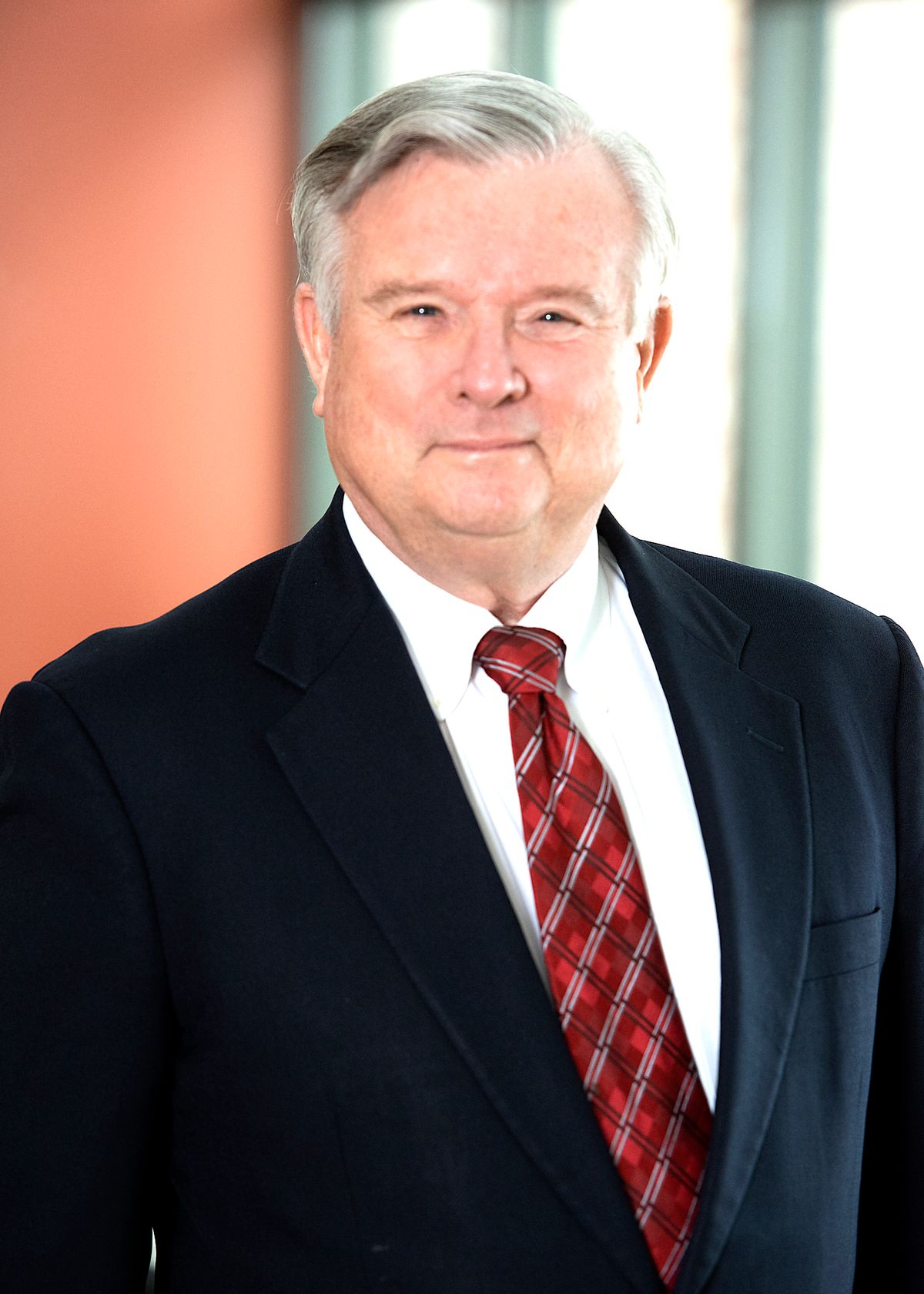
James Wilson directs the Orphan Disease Center at the University of Pennsylvania, where he explores gene therapy approaches to treat rare genetic conditions.
credit: University of Pennsylvania
For many rare genetic diseases, researchers have engineered mouse models housing the underlying mutations. Even then, possessing the same genetic mutations as the patient might not be enough to make a reliable animal model. “The question is do they have the same consequences clinically of that mutation?” said James Wilson, a gene therapy researcher and director of the Orphan Disease Center at the University of Pennsylvania. For example, mice featuring the genetic mutations present in cystic fibrosis do not show the same pulmonary manifestations of the disease (2).
Sometimes, researchers stumble upon a naturally occurring model of a rare disease rather than needing to engineer one. In the late 1970s, veterinarians were puzzled when they saw a cat named Rosebud who was losing the ability to walk. They referred him to scientists at the University of Pennsylvania who discovered that Rosebud had the disease known in humans as Hurler syndrome, the most severe form of mucopolysaccharidosis type 1 (MPS 1) (3,4). Soon after, they discovered the same disease in dogs (4). Approximately one in 100,000 people are born with Hurler syndrome due to mutations in the gene encoding α-L-iduronidase, a lysosomal enzyme that breaks down a polysaccharide, causing toxic accumulation that impairs cognitive and motor function (5). The affected cats and dogs sufficiently reproduce the anatomy of the human central nervous system and the neurological effects of Hurler syndrome, providing a valuable model for testing drug distribution and efficacy (6).
To identify a biomarker for the disease, Wilson’s team analyzed the cerebrospinal fluid of the Hurler syndrome dogs and found that it showed elevated levels of a polyamine molecule (7). They then studied patient samples and observed that the concentration of this polyamine correlated with the severity of the disease. Undergoing a bone marrow transplant to generate cells with the normal enzyme reduced the amount of the molecule, suggesting that it could be measured to evaluate new treatments.
Building on this foundation, Wilson’s group is developing a vehicle to deliver the wild type gene for Hurler syndrome to the human brain — just one of their many gene therapy projects. “Gene therapy has the potential to transform the landscape for patients with rare diseases because most of them are due to single gene defects,” Wilson said. “It has become the most extensively developed approach for treating rare diseases because of the simplicity of its concept. You have a defective gene, and as a result, you have this disease, so we'll put the normal version of the gene in.” While the genetic sequence, level of gene introduction, and delivery location need to be optimized for each disease, gene therapy provides a powerful tool to treat the root cause of many rare diseases.
Sharon Barr and her colleagues at Alexion, AstraZeneca Rare Disease have developed inhibitors for complement proteins to block a dysregulated immune response that drives several rare diseases.
credit: Alexion, AstraZeneca Rare Disease
Researchers at Alexion, AstraZeneca Rare Disease have pioneered another generalizable platform for treating rare diseases in the development of inhibitors for the complement system. This network of protein pathways within the innate immune system can become dysregulated, damaging healthy cells and tissues. “We have learned that this is a biological mechanism that is present in more than one disease,” Barr said. “Based on our understanding of disease progression, biomarkers, and pathophysiology, we teach ourselves, together with our collaborators, which pathways we think are predominant drivers, and then we tailor our therapeutic within the complement pathway to match it to the disease of interest.”
One of the company’s inhibitors of a key complement protein is FDA-approved for four diverse rare diseases: a blood disease, a kidney disease, and two neurological diseases. “We don't need to invent a brand new drug every time,” Barr said. “And that's good, because it takes a long time to invent a new drug, and patients are waiting.”
Barr keenly appreciates this sense of urgency; her own daughter survived a rare form of cancer, and her son recently received an effective drug for a rare autoimmune disease. “You would think, ‘How surprising that somebody who works in rare disease would have two children with rare diseases,’ but not really, because statistically, 10 percent of us are carrying the burden of a rare disease,” she said. “As I talk to my colleagues, statistically, you shouldn't be surprised to know that so many of us are facing a rare disease or managing one for our loved ones. And I think there's a reason why we collectively find our work so meaningful and rewarding. It's because we're creating solutions for families like our own.”
Old and improved
Just as the complement system mediates multiple rare diseases, a biological pathway that represents a drug target for a common disease might also be involved in a rare disorder. Researchers are therefore investigating if existing approved drugs may be repurposed to treat rare conditions.
Reena Kartha, a molecular pharmacologist at the Center for Orphan Drug Research at the University of Minnesota, explores repurposed drugs to treat Gaucher disease. This rare disorder is caused by an abnormality in the lysosomal glucocerebrosidase enzyme that leads to a harmful buildup of lipid substances in the liver, spleen, and bone marrow. The standard treatment involves injecting the normal enzyme, but “you're not addressing the defective protein that is hanging around in the cells in [the patient’s] body, and that can cause a milieu of other symptoms,” Kartha said. “There are patients who have been on treatment for more than 20 years, and they still have ongoing issues with pain and fatigue.”
Suspecting that the lingering defective enzyme triggers oxidative stress, Kartha collected patient blood samples and identified biomarkers of oxidative stress and inflammation that were elevated in people with type 1 Gaucher disease, the mildest and most common form (8). She is now conducting a clinical study to evaluate the effects of N-acetylcysteine, an approved antioxidant dietary supplement, on oxidative stress by measuring blood biomarkers, acquiring brain images to analyze metabolite levels, and incorporating patient-reported experiences of pain and fatigue (9). Using an approved drug allowed her to bypass many of the preclinical hurdles needed to test it in humans, which is especially handy as there is no animal model for type 1 Gaucher disease. The goal is to determine if N-acetylcysteine could provide a beneficial adjunctive therapy to the enzyme replacement regimen.
James Cloyd directs the Center for Orphan Drug Research at the University of Minnesota, where he applies pharmaceutical principles to develop drugs for rare seizure disorders.
credit: James Cloyd/University of Minnesota
Other researchers are improving upon drugs with known activity toward rare diseases. James Cloyd, a clinical pharmacologist and director of the Center for Orphan Drug Research, investigates better ways to deliver benzodiazepine drugs, which regulate neurotransmitter activity to treat rare seizure disorders. Intravenous administration of these drugs is challenging in the out-of-hospital settings where seizures spontaneously occur, so Cloyd developed a rectal benzodiazepine gel. “It works really well, except how many people do you know who want their pants pulled down at the mall to get this treatment?” Cloyd said. “So, we immediately began to look at an alternative.”
The new plan was nasal delivery, but this route presents its own limitations. Benzodiazepine drugs dissolve best in organic solvents, not water, which irritate and risk long-term damage to sensitive nasal tissue. Cloyd’s team developed a benzodiazepine prodrug, a modified, inactive version of the drug that’s highly soluble in water (10). At the time of administration, this molecule is mixed with an enzyme that converts it to the active drug, forming a super concentrated solution that rapidly absorbs across the nasal membrane and into the brain. “If we're successful with the prodrug, that really would represent a new chemical entity,” Cloyd said. “We built on a prior compound, but basically, you would be taking that from just about the very beginning of drug development.”
For Cloyd, the motivation to develop drugs for orphan diseases comes from the direct impact he has witnessed for patients. A mother approached him at an event to tell him that the rectal benzodiazepine gel, despite any embarrassing mall incidents, was life-changing for her family. “That’s all you need,” Cloyd said. “Using that as a trigger, I could see how focusing on rare disorders, particularly neurological conditions, and applying some of the tools not only that I have, but bringing in other people, really great colleagues, we can help out and make lives better.”
Joining voices
For some rare diseases, the major barrier to drug development isn’t designing the drug itself but evaluating its effect on the course the disease takes throughout an individual’s lifetime. “For rare diseases, oftentimes, there's not enough known about the natural history to design a proper clinical trial because you have to select, in most study designs, a single primary endpoint on which the trial hinges. How do you pick the right one if you don't know very much about the disease or its rate of progression or its main features?” said Edward Neilan, chief medical and scientific officer of the National Organization for Rare Disorders (NORD), a nonprofit organization founded by the same grassroots protestors at the White House 40 years ago.
Assembling enough patients to conduct longitudinal studies and clinical trials is challenging when so few people live with a specific disease, Wilson said. For genetic diseases caused by a single gene defect, between two-thirds and three-quarters of affected individuals reside beyond the United States. “We have to broaden our horizon outside of the US, not only to make sure we gain access to enough patients, but we also want to make sure that any advances that happen are distributed globally so that there's global access and global impact,” Wilson said.
We have to broaden our horizon outside of the US, not only to make sure we gain access to enough patients, but we also want to make sure that any advances that happen are distributed globally so that there's global access and global impact.
- James Wilson, Orphan Disease Center
Researchers at the Orphan Disease Center initiated a study of individuals across the world with Lesch-Nyhan disease, an extremely rare and severe neurological disorder caused by a single gene defect that affects approximately 1 in 300,000 people (11). Using a specialized rare disease data platform, the team is compiling their medical records and analyzing motor function and behavioral symptoms in people of different ages to construct a picture of the evolution of the disease over time.
To enable similar studies for the entire spectrum of rare diseases, NORD has launched the IAMRARE® registry program, in which patients complete surveys about their experiences living with rare diseases (12). More than 13,000 people representing approximately 40 rare diseases have participated in the registry to date. At its most basic level, the registry helps identify patients with a rare disease that may enroll in a clinical trial. “But the real desire of the IAMRARE® platform is to do patient led natural history studies,” Neilan said. While there are about 7,000 rare diseases, the pharmaceutical industry has focused on only 100 or 200 of them, according to Neilan. “That uneven attention is another problem that we’re trying to solve.” By building a better understanding of how a disease develops in the absence of a drug intervention and guiding the design of clinical trials, Neilan hopes that information from the registry will help drive research into rare diseases that pharmaceutical companies have thus far neglected.
The Rare Diseases Clinical Research Network (RDCRN) through the National Institutes of Health (NIH) shares the goal of laying the groundwork necessary to prepare rare disease indications for clinical trials (13). Researchers and patient advocates, who are few and far between for each individual rare disease, come together to form consortia for related rare diseases. The 20 consortia work together to determine the “who, what, where, when, and how” of clinical trial design, said Tiina Urv, the director of the RDCRN. For example, the researchers may uncover a biologically distinct subpopulation of a disease, allowing a drug to be tested in a more targeted cohort.
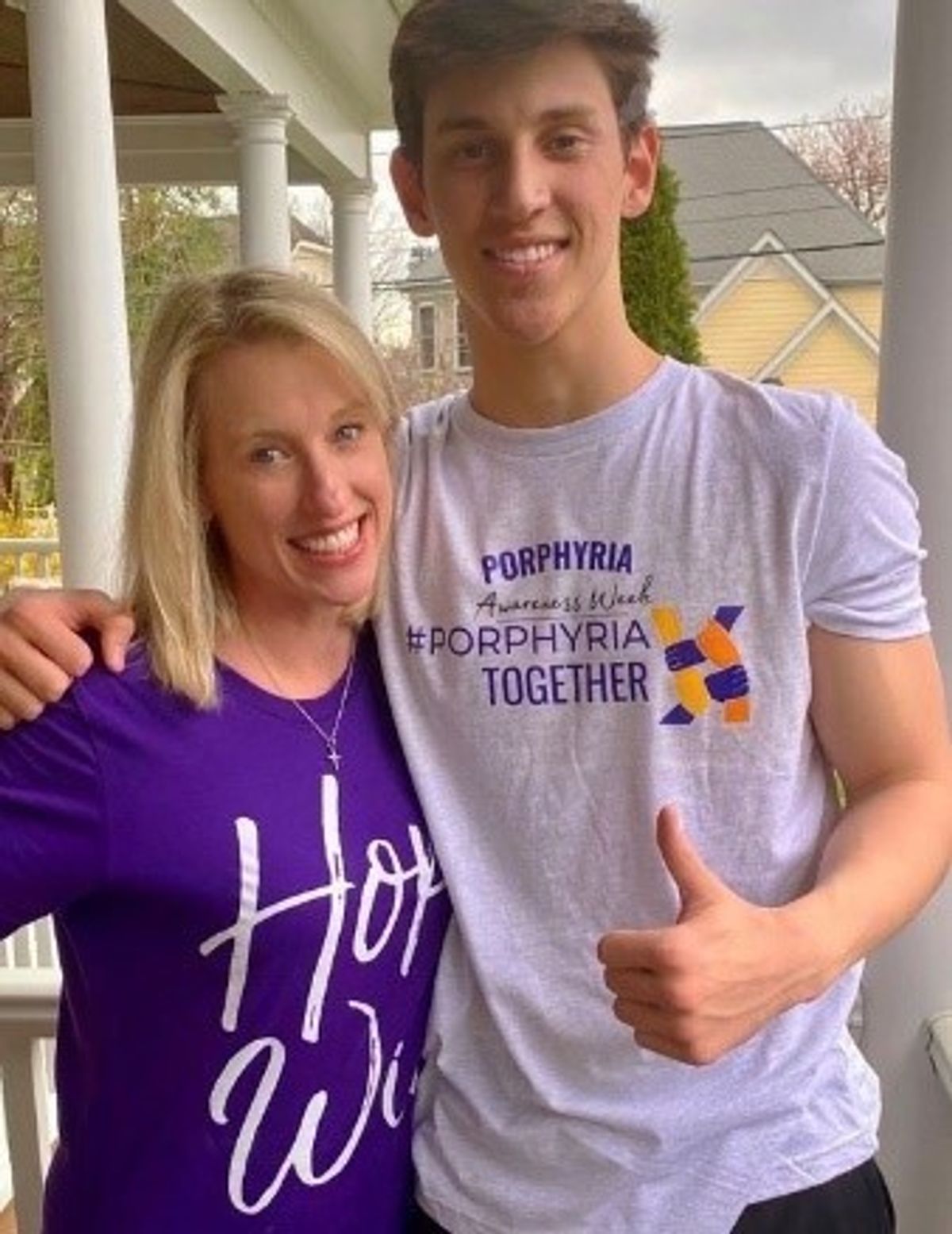
Kristen Wheeden serves as a patient advocate for the Rare Diseases Clinical Research Network’s porphyrias consortium, where she communicates the experience of her son Brady and others living with porphyria to inform the design of clinical studies.
credit: Kristen Wheeden
The patient advocates also play a critical role in developing meaningful and reasonable studies. “We’re not paying that popular trend lip service,” Urv said. “They’re expected to be at the table with the investigators looking at how the studies are designed.” For example, the patient advocates may provide feedback on how feasible it would be for a patient to lie still in an MRI machine or travel to a study site. “If a clinical trial fails, you want it to fail because the drug doesn't work, not because you have the wrong patients in the study, not because your patients are all dropping out of the study because they don't tolerate your measures,” Urv said.
Kristen Wheeden serves as a patient advocate for the consortium for porphyrias, a group of rare disorders caused by irregular production of the heme group in hemoglobin. Her son Brady lives with erythropoietic protoporphyria, a phototoxic form that causes him burning pain after about a minute of sun exposure. Clinical trials for erythropoietic protoporphyria have been hindered by the lack of an outcome measure by which to evaluate the efficacy of a drug. Drawing on her son’s experience and her work as president of the United Porphyrias Association, Wheeden collaborated with researchers in the consortium to find a way to use an early warning signal of sun-induced pain as a safe and reliable efficacy outcome measure (14).
“Alone, someone is rare, but when you pool people together, within even a specific type of porphyria, you get a larger voice. And when you join those voices, you really understand the patients, the diseases, their struggles, where they would like to see potential improvement, and what would be a good impact for them,” Wheeden said. “I see the RDCRN as really advancing the effort of greater research and therapeutics in rare disease through bringing together a network of physicians, researchers, and patient leaders and together focusing on the tough issues that face each group of diseases.”
Urv drew an analogy to curling. “I always think that the scientists and the patients are throwing the stone, and my responsibility is to sweep to try to help make things go a little more smoothly for them,” she said. “I see the NIH’s job as really doing what we can to help smooth or ease the road to developing treatments.”
A hopeful future
In addition to conducting research to facilitate drug development, many of these institutions are involved in public policy, education, and outreach efforts to advocate for rare disease treatment. NORD continues to defend the Orphan Drug Act against current legislative threats and has organized Rare Disease Advisory Councils comprised of stakeholders who provide recommendations that help shape state policies on rare disease (15). “You need the public policy part to make sure that the deck, which is inherently stacked against rare diseases, can continue to have some incentives,” Neilan said. “If we didn't have active advocacy to keep a finger on the scale and make it financially attractive for a biotech or pharmaceutical company to work on a rare disease that has only a small number of potential customers, we wouldn't be getting any of this development.”
Reena Kartha investigates approved drugs as adjunctive therapies for rare genetic diseases and teaches students at the Center for Orphan Drug Research about orphan drug development.
credit: Reena Kartha/University of Minnesota
At the Center for Orphan Drug Research, Kartha created a course for undergraduate students called Rare Diseases: What it Takes to be a Medical Orphan (16). The course covers topics such as rare disease models, natural history studies, clinical trials, the orphan drug development process, and the Orphan Drug Act with the goal of exposing the next generation of researchers, policymakers, and healthcare providers to the rare disease field. “They need to think about this community who are not rare if you take them all together,” Kartha said. “Nobody was doing it. Somebody had to do it. And I just thought, I will do it.”
In the course, students worked with patients and patient advocacy groups to learn about the barriers to research and treatment development for a specific rare disease and created Tik Tok videos to share their findings. “If I had gone in there and given 16 lectures on a disease, I'm sure that as soon as the semester was over, they would have forgotten it,” Kartha said. “But they worked with the community for that project, and I realized that is something that they will remember forever.”
Researchers at the Center for Orphan Drug Research also collaborated with the theater and dance department at the University of Minnesota to produce a play called RARE: Stories of Dis-ease, which integrated science and the arts to raise awareness about the challenges of living with a rare disease (17). The play was performed in seven cities, and while the researchers didn’t appear on stage, they served as scientific advisors. They gave a condensed Rare Disease 101 lecture to teach the performers the basics behind orphan drug development and brought in people who could discuss their experiences with rare diseases from different perspectives, including patients, physicians, and caregivers. After watching rehearsals, Cloyd helped the playwrights make a few important tweaks to the script. “I suggested a couple of things that they might modify,” he said, “including that they should emphasize at the end of the play that there's hope.”
References
- National Organization for Rare Disorders. Orphan drugs in the United States: an examination of patents and orphan drug exclusivity. At <https://rarediseases.org/wp-content/uploads/2021/03/NORD-Avalere-Report-2021_FNL-1.pdf>.
- Lavelle, G.M., White, M.M., Browne, N., McElvaney, N.G., & Reeves, E.P. Animal models of cystic fibrosis pathology: phenotypic parallels and divergences. Biomed Res Int 5258727 (2016).
- Haskins, M. E., Jezyk, P.F., Desnick, R.J., McDonough, S.K., & Patterson, D.F. Alpha-L-iduronidase in a cat: a model of mucopolysaccharidosis I. Pediat Res 13, 1294-1297 (1979).
- Haskins, M. et al. Animal models for mucopolysaccharidoses and their clinical relevance. Acta Paediatr Suppl 91, 88-97 (2002).
- Sakuru, R. & Bollu, P.C. Hurler syndrome. StatPearls: Treasure Island, 2022.
- Hinderer, C. et al. Neonatal tolerance induction enables accurate evaluation of gene therapy for MPS I in a canine model. Mol Gen Metab 119, 124-130 (2016).
- Hinderer, C. et al. Abnormal polyamine metabolism is unique to the neuropathic forms of MPS: potential for biomarker development and insight into pathogenesis. Hum Mol Genet 26, 3837-3849 (2017).
- Kartha, R.V. et al. Patients with Gaucher disease display systemic oxidative stress dependent on therapy status. Mol Gen Metab 25, 100667 (2020).
- Kartha, R.V. et al. Preliminary N-acetylcysteine results for LDN 6722 - role of oxidative stress and inflammation in Gaucher disease type 1: potential use of antioxidant anti-inflammatory medications. Mol Gen Metab 126, S82 (2019).
- Cloyd, J., Haut, S., Carrazana, E., & Rabinowicz, A.L. Overcoming the challenges of developing an intranasal diazepam rescue therapy for the treatment of seizure clusters. Epilepsia 62, 846-856 (2021).
- Nanagiri, A. & Shabbir, N. Lesch Nyhan syndrome. StatPearls: Treasure Island, 2022.
- National Organization for Rare Disorders. IAMRARE® program powered by NORD. At <https://rarediseases.org/advancing-research/patient-registry-program/>.
- National Institutes of Health. Rare Diseases Clinical Research Network. At <https://www.rarediseasesnetwork.org>.
- Wensink, D. et al. Erythropoietic protoporphyria: time to prodrome, the warning signal to exit sun exposure without pain-a patient-reported outcome efficacy measure. Genet Med 23, 1616-1623 (2021).
- National Organization for Rare Disorders. Rare Disease Advisory Councils. At <https://rarediseases.org/rare-disease-advisory-councils/>.
- Center for Orphan Drug Research. Rare Diseases: What it Takes to Be a Medical Orphan. At <https://www.pharmacy.umn.edu/centers-and-institutes/center-orphan-drug-research/research-education-opportunities/what-it-takes-to-be-a-medical-orphan>.
- Center for Orphan Drug Research. RARE: Stories of Dis-ease. At <https://www.pharmacy.umn.edu/centers-and-institutes/orphan-drug-research/rare-disease-day/rare-disease-theater-project>.