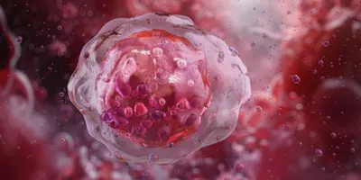
To successfully infect a host, the bacteria Listeria monocytogenes rely on their ability to invade cells, multiply in them, and subsequently leave. These intracellular pathogens invade many kinds of human cells, including macrophages.

Now, a team of researchers led by Carine Smith, a physiologist from Stellenbosch University, co-opted L. monocytogenes proteins to help macrophage-drug carriers release drugs at the appropriate site and deliver more potent drugs with fewer off-target effects (1).
Macrophage-based drug delivery can allow for smaller doses by preventing degradation during transit, and their shielding effect can mean more toxic pharmaceuticals can be used with lower risk. “It provides a means by which we can address drug-resistant and chronic conditions with little risk to the patient,” said Smith. However, in many cell-based drug delivery systems, drug delivery relies on passive diffusion to release the drugs, or because drugs often become trapped within vacuoles in the cell, the cell must die to release the drug. Smith’s team turned to intracellular bacteria to solve this problem.
“[Intracellular bacteria] have these nifty mechanisms by which they manipulate the cell membrane to make almost like little straws through which they can escape when they're ready to disseminate to a different area,” said Smith.
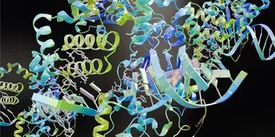
For L. monocytogenes, this escape occurs in a two-step mechanism orchestrated by many proteins including listeriolysin-O (LLO) and actin assembly-inducing protein (ActA). First, LLO creates holes in the phagosome membrane so that the bacterium can enter the cytosol of the macrophage. Then, to exit the macrophage, ActA induces actin polymerization which is necessary to leave the host cell.
Smith and her team first expressed LLO and ActA fused to green fluorescent protein (GFP) and purified them. Then, they coated polystyrene beads with these proteins to create what Smith called a “synthetic microbe” and introduced them into macrophages. Because of the GFP tag on the synthetic microbe, they could track their location.
Using several microscopy techniques including super-resolution structured illumination microscopy, confocal microscopy, and imaging flow cytometry, Smith and her team tracked the macrophages’ physical changes as well as the internalization and location of the beads. Compared to macrophages exposed to control serum-coated beads, macrophages exposed to the synthetic microbes did not accumulate beads, but spikes in their cell membranes appeared, indicative of actin polymerization. This suggested that the synthetic microbe exited the cell. The synthetic microbes also accumulated towards the periphery of the macrophages, a characteristic similar to what L. monocytogenes do inside infected cells. In contrast, control beads were more evenly distributed in the macrophage.

“Our approach is unique in that the cells will survive afterwards and play a role in resolution of inflammation, clearing the tissue, and getting it ready for regeneration, which is a much-desired outcome after you've delivered your drug,” said Smith.
“We know that macrophages are attracted to disease sites, but if they're carrying a therapeutic agent, you can imagine that therapeutic agents could be a lot more effective if the macrophage is acting as a deliverable for these agents,” said Howard Gendelman, an immunopharmacologist and microbiologist at the University of Nebraska Medical Center who was not involved in the study.
It provides a means by which we can address drug-resistant and chronic conditions with little risk to the patient.
- Carine Smith, Stellenbosch University
“The idea of better packaging these agents, to homing these agents, to facilitating the activity of these agents, are new and exciting opportunities for how a cell-based system can actually increase the delivery of antimicrobial or therapeutic agents,” said Gendelman.
Smith is now optimizing delivery conditions using zebrafish larvae infused with human immune cells. She hopes this model can help them understand how to control when drugs get released or not.
“[Macrophage-based drug delivery] holds promise for people who have been struggling with hard-to-heal issues,” said Smith. “We're very excited about the idea that a macrophage can enter even bone tissue, areas which are really hard to reach with conventional medication that's just put in the blood supply or in circulation.”
Reference
- Van Staden, A.D.P. et al. Harnessing Microbial Effectors for Macrophage-Mediated Drug Delivery. ACS Omega 9, 18260–18272 (2024).