Scientists frustrated by the current approach to drug development are joining biotech start-ups focused on developing therapies targeting a long-forgotten organelle: the condensate.
Condensates are membraneless organelles formed by the concentration of collaborative biomolecules such as transcription factors and DNA. The first condensate was described in 1835 and dubbed the nucleolus since it speckled the nucleus (1). Although we know now that the nucleolus is a liquid filled bag of translational machinery, including rRNA and protein, and the site where protein pumping machines known as ribosomes are made and assembled, no one paid the nucleolus much attention until 1950, when an understanding of its function started to emerge. The term “condensate” wasn’t coined until nearly 60 years later.
In 2008 during a course at Cold Spring Harbor Laboratory, Cliff Brangwynne, a biophysicist at Princeton University, and his students noticed that RNA in fertilized worm embryos aggregated and dissociated into droplets; it was like looking into a cellular lava lamp. These condensates, now known as P granules, which contain specific germline RNA in worm eggs, inspired Brangwynne to pen a letter to Science about how P granules might form via a phenomenon called liquid-liquid phase separation (LLPS), where biomolecules like rRNA and proteins coalesce into a liquid like droplet resembling wax in a lava lamp (2).
Richard Young is a condensate researcher at the Massachusetts Institute of Technology (MIT) and co-founder of Dewpoint Therapeutics.
CREDIT: RICHARD YOUNG
“It’s not a crazy new idea; it just wasn’t deployed in biology. It’s actually a property of polymers, and remarkably, biologists like me had simply not studied that fundamental polymer chemistry and physics that would have told us that the three key biopolymers, DNA, RNA, and protein tend to form these condensates. It’s a basic property of matter that math and physics would predict,” said Richard Young, a condensate researcher at the Massachusetts Institute of Technology (MIT) and co-founder of Dewpoint Therapeutics.
But few others felt inspired by the application of this basic concept in physics, at least at first. In 2011, Brangwynne published a more detailed description of how LLPS may drive the formation of condensates (3). And the next year, Michael Rosen, a biophysicist at the University of Texas Southwestern Medical Center, reported in Nature that his team had replicated these proteinaceous lava lamps in a test tube by using signaling proteins (4).
A flurry of research followed, suggesting that condensates have their hand in everything from transcription to neuronal signaling. Growing evidence even shows that drugs can’t always penetrate condensates to reach targets that reside inside, which decreases their efficacy (4). And sometimes drugs concentrate in the wrong condensate, causing unwanted side effects. New biotech companies like Dewpoint Therapeutics and Nereid Therapeutics have emerged in the past five years to design better drugs that localize to the right condensate.
“I firmly believe that targeting condensates is going to change the way we think about developing drugs and really change the way we think about human disease,” said Isaac Klein, chief scientific officer of Dewpoint Therapeutics.
Defining a condensate
Klein became disenchanted with the cancer drug discovery process after beginning his oncology fellowship about five years ago. He noticed that most drugs were designed for a small subset of patients with a specific mutation. He wondered if there was a way to design drugs that targeted a more conserved disease mechanism across more patients.
“When I heard about condensates, I realized that they provide a way of attacking the most important undruggable targets. And they allow us to think about how the cell integrates multiple signaling abnormalities into one,” said Klein. “It’s exactly the opposite of how all cancer biologists and the entire cancer drug discovery community think about developing drugs. I thought ‘wow, that’s a much more powerful approach to help cancer patients than any other.'
Klein decided to leave the clinic to join the academic world to study condensates. He joined Young’s lab and soon published a paper in Science that reported that some cancer drugs localize in nuclear condensates, which can positively or negatively affect their efficacy (4).
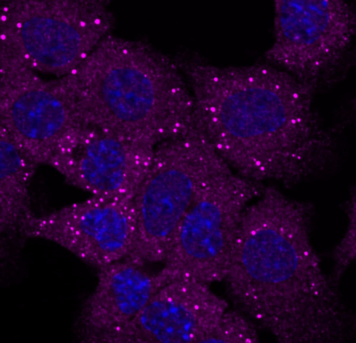
Cells are decorated with droplet-like condensates (dark purple).
Credit: dewpoint therapeutics
In this study, Klein and Young tagged two chemotherapeutic drugs, cisplatin and tamoxifen, so that they could watch how the drugs interacted with condensates composed of transcription factors and DNA.
Cisplatin accumulated in condensates that contained the transcriptional coactivator, Mediator Complex Subunit 1 (MED1), while it simply diffused through other transcriptional condensates. Cisplatin, which kills cancer cells by binding DNA and blocking transcription, was more active in MED1-containing condensates as well.
The researchers found a biological basis for cisplatin’s preference. MED1 contains many aromatic amino acids, which possess a type of carbon arrangement that forms a decentralized ring of electrons. These aromatic amino acids attracted other molecules, including cisplatin, that contain aromatic rings, exemplifying how drugs could be chemically modified to preferentially accumulate in a particular condensate. This could make drugs more effective and limit unintended side effects wreaked by unsuspecting small molecule drugs stumbling into the wrong condensate.
Young and his team also discovered how a common cancer mutation may prevent tamoxifen from effectively localizing in its target-containing condensates. Tamoxifen targets a protein commonly overexpressed in breast cancer, estrogen receptor alpha (ERa), for degradation. The researchers discovered that when tamoxifen enters condensates containing ERa, it evicts the protein. But when an ERa protein containing a mutation known to cause tamoxifen resistance was present in the condensate, tamoxifen was evicted instead. Tamoxifen then concentrated into MED1- containing droplets which, unlike cisplatin, made the drug less effective.
This landmark study is part of the basis of Dewpoint Therapeutics’ strategy to create new drugs and manipulate existing drugs to target the right condensate.
“Now that we know that small molecules can distribute within cells in a way that they either simply passively transfer through condensates, they’re excluded, or they concentrate, we can modify small molecules so that they have this selective property of concentrating in the condensate where their target lies,” said Young.
Designing drugs with condensates in mind
Dewpoint Therapeutics isn’t only interested in cancer. In addition to oncology, they are investigating drugs in neuromuscular disease, cardiac disease, and virology. They’ve teamed up with several well-established pharma companies like Pfizer, Merck, and Bayer to accomplish their lofty goals. They’ve developed a broad platform to discover which diseases are condensate related and what malfunctioning condensates could be targeted in disease.
“Many different mutations and expression level changes in seemingly disparate proteins can all impact the function of one condensate. So, by addressing a single condensate, rather than by addressing the many alterations that occur in different proteins, you can potentially address a much greater patient population with one molecule, rather than dividing it up into a whole bunch of subpopulations and then pursuing a drug discovery campaign for those individually,” said Klein.
Dewpoint Therapeutics is developing condensate-directed drugs for amyotrophic lateral sclerosis (ALS). Although many people with ALS have different disease-causing mutations in a variety of proteins, 97% of patients have mislocalized aggregates of the RNA processing factor known as trans-activating response element DNA-binding protein of 43kDa (TDP43) in some of their neurons (6). This aggregation affects the function and formation of TDP43-containing nuclear condensates (7).
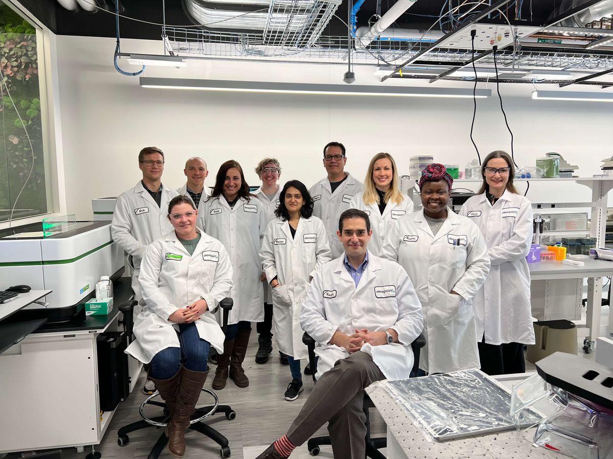
Isaac Klein, the chief scientific officer at Dewpoint Therapeutics, and his team aim to make better drugs by targeting condensates.
CREDIT: DEWPOINT THERAPEUTICS
“By targeting the condensate, you can treat all of the patients regardless of the etiology of their disease. This is in direct contrast to the more common, more predominant drug discovery strategy which is ‘oh, they have a mutation, let’s target that.’ That’s predominantly failed,” said Klein.
Dewpoint Therapeutics isn’t the only new biotech company with their eyes on condensates. Other companies such as Faze Medicines, Transition Bio, and Nereid Therapeutics have emerged in the last couple of years and are gearing up to develop condensate-targeted therapies, each with their own unique spin.
“Most of our competitor companies are either very focused on particular disease areas or technologies and haven’t taken this very broad platform approach,” said Klein.
Nereid Therapeutics takes a more technology-based, physics driven approach. The company was co-founded by Brangwynne, who identified and established LLPS as a driving force behind condensate formation.
“The approach that we are taking is a very biophysically based approach. Some folks in the field are taking a more descriptive approach whereas our approach is more quantitative,” said John Reilly, chief scientific officer of Nereid Therapeutics.
Spiros Liras, chief executive officer of Nereid Therapeutics said that their approach really is a “marriage of physics with cellular biology.” They plan on using optogenetics to visualize the formation of condensates through LLPS, which will allow them to understand how this process is affected in disease and how therapeutics can target this physics-driven process. (It will also produce the ultimate biological lava lamp.)
“It actually allows us to visualize the formation of these droplets and consider what their function is, and what quantitatively influences it. It’s a different way to actually do drug discovery. It’s not really about targeting a specific target with inhibition or activation. It’s more about understanding what happens in the reaction in the cell,” said Liras.
Gary Karpen, a condensate biologist from the University of California, Berkeley who is not involved in any of the new condensate biotech start-ups is excited to see how these new companies will move condensate research from the bench to the clinic.
“The issue is, what is it you're trying to target? Is it a good thing to target the solution of this particular condensate? All the companies need to demonstrate that they can find ways to manipulate formation or dissolution of these condensates in a way that's useful for disease therapeutics. That's going to be challenging,” said Karpen.
Liras thinks that it can be done. He views the other biotech start-ups popping up left and right as fellow travelers on a journey to bring condensate-based therapeutics to patients, although they are taking different paths.
“It’s reassuring that there’s not just one company in it, and yet the field has not grown to the point yet where it’s saturated. I think there’s a lot of opportunity for all the companies to bring to the forefront what is a novel approach for drug discovery,” said Reilly.
Check out this infographic to learn more about condensate formation.
References
- Pederson, T. The Nucleolus. CSH Perscpect Biol 3, a000638 (2011).
- Brangwynne, C.P. et al. Germline P Granules Are Liquid Droplets That Localize by Controlled Dissolution/Condensation. Science 324, 1729-1732.
- Brangwynne, C.P. et al. Active liquid-like behavior of nuceoli determines their size and shape in Xenopus laevis oocytes. PNAS 108, 4334-4339 (2011).
- Li, P. et al. Phase Transitions in the assembly of multi-valent signaling proteins. Nature 483, 336-340 (2012).
- Klein, I.A. et al. Partitioning of cancer therapeutics in nuclear condensates. Science 368, 1386-1392 (2020).
- Schmidt, H. B. et al. Phase separation-deficient TDP43 remains functional in splicing. Nat Comms 10, 4890 (2019).
- Altman, et al. Axonal TDP-43 condensates drive neuromuscular junction disruption through inhibition of local synthesis of nuclear encoded mitochondrial proteins. Nat Comms 12, 6914 (2021).