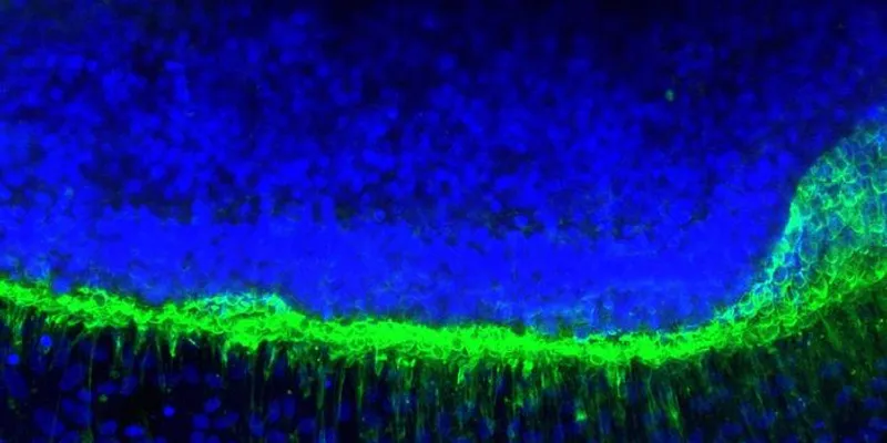
In the most common form of Batten disease, known as CLN3 disease due to mutations in the CLN3 (ceroid-lipofuscinosis, neuronal 3) gene, the first symptom is usually loss of vision in early childhood. “It often precedes neurological decline by years, and it's the one that most immediately changes a child's ability to read, learn, and move through the world independently,” Ruchira Singh, a retinal and neurodegenerative researcher at the University of Rochester, told DDN. A rare, fatal inherited disorder of the nervous system, CLN3 disease typically begins in childhood and causes progressive mental and physical decline.
Scientists have not fully understood what causes this vision loss, in large part because previous human in vitro models were not able to represent the spatial organization between photoreceptors and retinal pigment epithelial (RPE) cells in the eye. But that changed recently when Singh’s lab developed a new 3D retinal organoid model derived from human pluripotent stem cells. The new model successfully “reproduces the earliest and most consistent pathology seen in CLN3 disease patients, the disorganization and loss of photoreceptor outer segments,” said Singh.
The new way of studying the disease allowed the researchers to move beyond studying the disease as neurodegenerative in nature and show that the mutation in RPE cells — which form a single layer to protect and support photoreceptor nerve cells — is enough to cause photoreceptor degeneration on its own.

“Our data show that the pathological driver of neuronal degeneration can be non-neuronal and non-cell autonomous. We show that if the RPE cells fail first, photoreceptors will degenerate consequently,” Singh explained. “That reframed how we understood the disease.”
The findings from the 3D retina model also helped the team identify and test a new treatment to target the RPE cells in the organoid and a large animal model, which could lead to much better outcomes for patients one day.
How the organoid offers a better view
Singh’s team was excited to discover that their new 3D retina model better represented what happens in children with the disease. “We were reproducing, in a dish, the outer retina pathology that children with this disease experience and that mouse models had never been able to capture,” she said.
Their excitement grew further when they tested a novel treatment approach. The 3D retina model allowed them to see that reduced acid ceramidase levels acted as a molecular driver of photoreceptor degeneration, so they reasoned that enzyme replacement with recombinant human acid ceramidase (rhAC) might help. In both the 3D retina model and in a miniswine model, rhAC led to improvements in cellular health of the photoreceptor cells.
“Seeing a therapeutic target we identified in a dish hold up in a living animal is the part every one of these projects is working toward, and it doesn’t always happen,” said Singh.
“That reframed how we understood the disease.”
—Ruchira Singh, University of Rochester
After more preclinical testing, Singh and her team are hopeful that this new therapeutic approach could move into patients, especially because rhAC has already been studied for over a decade as an enzyme replacement therapy in Farber disease. Plus, its use in CLN3 disease to target vision loss might be even easier with fewer side effects. “We would deliver it intravitreally rather than systemically. That avoids the problem of getting a large enzyme across the blood-retina barrier, and intravitreal injection is already routine in ophthalmology,” said Singh.
Yet, before testing can begin, Singh’s team will work on optimizing the intravitreal rhAC in their miniswine model and determine the dose that could lead to a longer duration therapeutic.
“These are tractable questions, but they are ahead of us, not behind us,” said Singh. “We are actively seeking funding support to address this immediate and urgent gap, because it is the step standing between the mechanism we have identified and treatment that could reach patients.”
Continued success for organoid models
The use of the 3D organoid model was crucial to the team’s novel discoveries and offers another example of how this approach is advancing basic knowledge in neuroscience.

“The ability to have defined combinations of mutant and control cell types in a 3D human retina cell model allowed us to investigate whether RPE dysfunction alone is sufficient for initiating photoreceptor degeneration; a question that can’t be answered in currently available animal models, where every cell carries the mutation, or in a 2D culture that lacks the photoreceptor-RPE interaction,” explained Singh.
The new investigative approach led the team to test out a treatment that moved beyond targeting photoreceptors only — a strategy that Singh notes will be necessary to achieve long-term therapeutic impact. The work could be integrated into other therapeutics already under study targeting vision loss in CLN3 disease like gene therapies and drugs that work on lipid metabolism by suggesting a need to target the RPE cells as well.
Moving forward, Singh’s team will use their new 3D retina model to directly compare the therapeutic effiacy of rhAC along with other potential treatments — a task that was not fully possible before.
“The development of the 3D retina model provided a thus far elusive human platform for mechanistic and therapeutic studies,” said Singh.