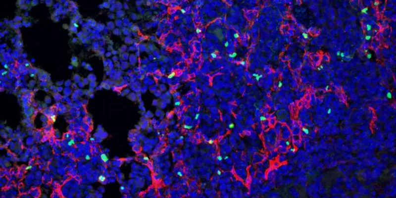
Imagine finding a ring of cells around the border of a tumor that look like normal cells, but they’re actually protecting the cancer within. That’s what researchers led by Olivia Ringham and Nicholas Arpaia at Columbia University Irving Medical Center recently found in new work published in Nature Immunology.
Using spatial transcriptomics to analyze what they thought were typical fibroblasts in a mouse model of lung cancer, the team found a population of immunomodulatory cancer-associated fibroblasts that express a gene called CHL1 (cell adhesion molecule L1-like) that regular fibroblast lung cells do not. The findings pointed to a potential new treatment strategy, which the researchers then tested in mice.
The discovery is one of the latest examples of how spatial transcriptomic analysis is driving progress in our basic understanding of how tumors adapt and evolve — including at the individual cell level — and consequently identifying new drug targets. “Single-cell transcriptomics has really had a huge development in theon the field over the last decade,” said Ringham in the press release. “It’s a great way to find hidden cell types and understand what those cells do.”
The global spatial transcriptomics market is now expected to grow from $495.2 million in 2026 to $1,306.4 million in the next seven years, with discoveries like the one from Arpaia’s team ultimately getting closer to improving patient care across all types of cancer.
Targeting the tumor’s protective mechanism
Because spatial transcriptomics allows scientists to view cells at precise spatial locations like the border of a tumor, Arpaia’s team was able to identify the seemingly-normal looking fibroblasts and study their activity in relation to the nearby lung tumor.
What they found was a sneaky way for the tumor to protect itself. The newfound cells recruited regulatory T cells, which are usually protective within the lungs as they keep the immune system from attacking every possible environmental antigen that enters. But in this context, the tumor recruits them as a sort of bodyguard around its perimeter, creating an immunosuppressive microenvironment that instead protects the cancer cells. The tumor extends its invitation through a signaling protein called CXCL9 (C-X-C motif chemokine ligand 9).
That discovery also opened up a new target for lung cancer. The researchers blocked the CXCL9 signaling system in the mouse model by inactivating the genes that code for it, which in turn decreased the amount of regulatory T cells that showed up at the border of the tumor — and allowed normal immune responses to get through and attack the cancer cells.
Importantly, the research team validated these findings in human lung cancer tissue samples and showed that these same types of fibroblast cells exist in human lung cancers, with more of them being associated with weakened immune responses against the tumor.
Future work could aim to figure out how these newly discovered cells arise in the lungs in the first place, since they are not found in the lungs of people without cancer. “Where do these cells come from? How are they formed?” said Ringham. “If we could stop that transformation, it could provide a great benefit.”