
Stephanie joined Drug Discovery News as an Assistant Editor in 2021. She earned her PhD from the University of California Los Angeles in 2019 and has written for Discover Magazine,...
Viewfull profile.Learn about oureditorial policies.
Some scientists get their start sorting fruit flies or picking bacterial colonies. Others dig up their dead pets every week to document their decomposition. Altos Labs cell biologist Alex Ritter is the latter.

Growing up in rural North Dakota, Ritter spent his days crawling around creeks and collecting animals he found there — everything from frogs and salamanders to snakes — much to the chagrin of his parents. While he recorded the decay of his deceased pet frog or disassembled fireworks to make new ones, Ritter didn’t realize that he was well on his way to becoming a scientist.
“I didn't really know what a scientist was when I was young,” he said. “I just saw what was on cartoons and that sort of thing. I didn't realize being a scientist was a job.”
But the first time he peered through the eyepieces of a microscope changed all that. Ritter discovered the world of cytotoxic T cells and their penchant for killing cancer and virus-infected cells. As a postdoctoral scientist at Genentech, Ritter used cutting-edge microscopy to visualize how cancer cells resist T cell killing, and now at Altos Labs, he’s ready to dive into how immune cells influence the process of aging and develop immune-based therapeutics.
How did you get interested in microscopy and cell biology?
I worked in a molecular biology lab at Carnegie Mellon University as a summer undergraduate student and spent most of the program cutting and pasting DNA on the bench to tag mitochondrial proteins with fluorescent markers. When I transfected my constructs into cells and finally went to the microscope, I was totally blown away. When we learn about mitochondria in textbooks, they look like beans that are full of spaghetti-like cristae. I expected to see little dots, but I saw a beautiful interconnected filamentous network. It was definitely a moment of wonder for me, and that's where I fell in love with microscopy and cell biology.
How did that experience lead you to immunology and cancer research?
It happened randomly. I was standing in front of my poster of my summer research at a cell biology conference, and a person came up to me and asked about my career plans. I had just been accepted into the National Institutes of Health (NIH) Oxford-Cambridge Scholars program. This person suggested that I reach out to her good friend named Gillian Griffiths at Cambridge who studies cytotoxic T cells. Griffiths was interested in using microscopes to study how killer T cells engage and destroy their targets, and I thought it was the coolest thing ever. Because the program is collaborative, I also worked with Jennifer Lippincott-Schwartz at the NIH who is a world-renowned cell biologist and who is especially famous for her work developing new microscope technologies.
What research questions did you work on with Griffiths and Lippincott-Schwartz?
I wanted to see the full process of what happens inside the killer T cell as it engages a cancer cell or virus-infected cell. There were a lot of fixed images and a few videos of that process. I developed new techniques to express constructs in cytolytic immune cells and to image them during the process of killing. I wanted to learn more about how the immune synapse — the place where the immune cell and its target cell meet — forms and the process of T cell killing. It was really an explosion of exciting, creative work because there were new microscope technologies that I could try, and we started getting amazing videos.
in one of the most detailed videos of this process ever captured.
CREDIT: ALEX RITTER
What was the most challenging part about capturing videos of immune and cancer cells?
T cells don't like to sit still for a picture, so when we put them on a surface, I had to figure out how to get them to adhere and crawl on glass. When it came time for imaging, I plated the cancer cells first and then sprinkled the T cells down. Often, I would find one T cell that looked like it was about to engage its target. I would start imaging, and it would stop or change direction. I realized after a while that T cells are photosensitive. If we don’t image them with gentle illumination, they don't behave normally. When I used extremely low levels of laser power in the microscope, they started to kill, and we discovered all sorts of interesting phenotypes.
How did you decide to investigate tumor cells’ resistance to T cell killing?
When I was making videos in my graduate work, I observed that sometimes cancer cells resisted killing, so when I went to Genentech for my postdoctoral research, I shifted my focus to study the target cell side. I found that the T cell often needed to secrete multiple rounds of toxic lytic granules to kill a cancer cell, and I wondered what was going on.
One of the toxins T cells secrete is perforin, which pokes holes in the membrane of the target cell. Another toxin, which goes through those little pores, enters the target cell and initiates a self-destruct sequence called apoptosis. In our videos, we saw that when T cells secreted perforin, the cancer cells seemed to repair their membranes really quickly. They basically fixed the holes almost as soon as the holes formed, and we wondered what was repairing them. I took a candidate approach and tagged proteins that could be involved in fixing wounds in plasma membranes while looking at their localization during T cell killing. I also started to knock them out to see if deleting any of them enhanced the ability of T cells to kill their targets. After this, we ended up with the ESCRT complex, which is important for a lot of different things in cells including membrane repair. Tumor cells recruited it strongly to the cell membrane, and when I knocked it out, we saw a big difference in the ability of T cells to kill tumor cells (1). That was super exciting for us.
CREDIT: ALEX RITTER
What was your reaction when you saw how quickly the ESCRT proteins rushed to the pore on the tumor cell?
If someone was standing by the microscope room, they would have heard me go, “Woohoo!” It was probably one o’clock in the morning when I saw that, but it was a result that I had been waiting to see for at least a year. I was in wonder at how fast it happened and how robust the result was. Every single cell that I imaged had a robust recruitment of ESCRTs. It's always nice when something is extremely repeatable because it gives us even more motivation to keep pushing forward.
What do you plan to do next?
If someone was standing by the microscope room, they would have heard me go, “Woohoo!” It was probably one o’clock in the morning when I saw that, but it was a result that I had been waiting to see for at least a year.
– Alex Ritter, Altos Labs
Working at Genentech has been amazing, but I want to reach into a different intellectual space. I will be joining a startup called Altos Labs. Although they aren’t working on cancer, they are working on a process that is indelibly linked to the immune system: aging. I won’t just be studying how the immune system responds to the aging body, but also investigating potential therapeutic interventions. We could potentially engineer immune cells to go into tissues or reengineer the tissues themselves. There's lots of space for cell therapy in the aging field, and I'm really interested in learning how we can potentially engineer these killer immune cells to make them useful in the context of aging and to cure diseases of aging.
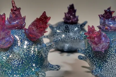
I also saw that you create glass blown art of immune cells. How did you get interested in that?
Honestly, the interest came in two ways. When I was a child, I was a bit of a pyromaniac. I used to take apart fireworks and rebuild them to make new fireworks. Also, years ago, I learned about the Blaschka family who created glass models of sea creatures in the mid-1800s. I was amazed at how realistic they could make these animals look out of glass, and I just thought, “Wow, if I can take what I see in the microscope and make it out of glass like that, then people could potentially connect with it better.” I’m a big fan of science communication, and I think finding interesting ways to engage people is so important.
How do you find that your immunology research informs your glass artwork?
When it’s hot enough, glass is a liquid. And cells, while they have structure, they're really liquid themselves. Instead of chiseling something out of stone, I’m working with a fluid to create a fluid, dynamic object. Cells are not colorful, but with glass, I can add vibrant colors and think about how to best represent what I see in the microscope using different colors and forms in glass.
I really hope my video microscopy and glass work can give cancer patients a visual representation of what's going to happen in their bodies when they get immunotherapy treatment. It's one thing to go into a doctor's office, get an injection of antibodies, and have the doctor say, “This is going to activate the immune system to destroy a tumor.” It's a whole other thing to see a killer T cell engaging a cancer cell and destroying it or to hold a physical glass piece of art depicting a T cell killing a cancer cell. I really believe in the power of positive thinking, and if people can imagine what’s happening inside of them, that can only help.
This interview has been condensed and edited for clarity.
Reference
- Ritter, A.T. et al. ESCRT-mediated membrane repair protects tumor-derived cells against T cell attack. Science 376, 377-382 (2022).

