Throughout her career, Jisook Moon, a neuroscientist at Cha University, has sought to develop stem cell-based treatments for neurodegenerative conditions like Parkinson’s and Alzheimer’s disease. About a decade ago, she tried injecting human stem cells directly into the veins of mice with Alzheimer’s disease symptoms, and she was pleasantly surprised to see improvements in cognition and reductions in neuroinflammation weeks after treatment (1). “Stem cells cannot go through the blood-brain barrier,” said Moon, so something else must have been contributing to the positive effect.
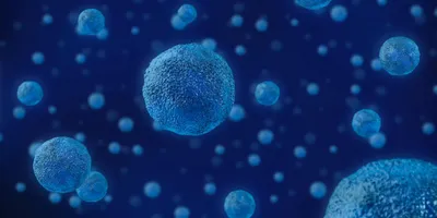
Her search for an explanation led her to extracellular vesicles (EVs) — the small, membrane-enclosed packets of molecules that cells throughout the body regularly release. Moon learned that EVs can cross the blood-brain barrier in both directions and communicate information about the health of otherwise inaccessible brain cells (2). “Then I realized that maybe EVs can be a tool for brain diagnosis,” she said.
Moon knew that she needed some way to distinguish brain-derived EVs from the other types present in any given blood sample. In a study published in Science Advances, she and her team identified the protein amyloid precursor-like protein 1 (APLP1) as a biomarker fit for that purpose (3).

Jisook Moon became interested in the diagnostic potential of brain-derived extracellular vesicles after witnessing their ability to cross the blood-brain barrier.
CREDIT: Jisook Moon
Around the time Moon first turned her attention to EVs, the protein L1 cell adhesion molecule (L1CAM) emerged as a candidate biomarker for distinguishing neuronal EVs and, ultimately, detecting brain disease (4). But in the years since, researchers including Moon have questioned how well that protein can fill the role. Tissues throughout the body express L1CAM, and forms of it are present in the blood unattached to EVs (5,6).
There are still researchers developing tools for isolating neuronal EVs using L1CAM as a biomarker (7). Victoria University molecular neuroscientist Andy Hill, who was not involved in the present study, doesn’t see any reason not to. “If you’re trying to diagnose a disease, if you do steps A, B, and C, and you get a marker that discriminates, that’s all that matters, right?” he said.
While those efforts unfold in other labs, Moon has decided to prioritize tissue specificity in her search for a better biomarker. At first, she thought she could find what she was looking for by running some proteomics or metabolomics analyses on blood samples. “But I failed,” said Moon. “The amount is so small I couldn’t do some sequencing.”
So, Moon searched through published databases and identified 35 proteins that are expressed exclusively in the brain and that are located in the region of the cell membrane where EVs form. When she searched for those proteins in the Brain RNA-Seq database, the one with the highest and most widespread expression throughout the brain was APLP1.
That protein emerging from Moon’s search didn’t come as a surprise to Hill. “A long time ago, I was working on APP, which is the amyloid precursor protein,” he explained. APP gets processed into the protein responsible for forming plaques in Alzheimer’s disease, and APLP1 came up in Hill’s work because of its structural similarity to the former. “When I saw the protein name come up in pulling down neuronal-derived EVs, I thought that made sense,” said Hill.
Moon confirmed that APLP1 was more specific to brain tissue than L1CAM with her own analyses of RNA and protein levels in various mouse organs. Then, she isolated EVs directly from mouse brain tissue using a separation technique Hill had previously developed and used a western blot to show that APLP1 was even more strongly expressed in the EVs than in the brain tissue as a whole (8).
Then I realized that maybe EVs can be a tool for brain diagnosis.
– Jisook Moon, Cha University
Satisfied that the protein could serve as the biomarker she was looking for, Moon developed an antibody and used it to pull APLP1-positive EVs out of mouse and then human blood plasma samples. She examined the contents of the EVs and confirmed that they carried proteins associated with brain tissue.
Moon plans to continue refining the antibody to ensure that it can isolate EVs from whole blood samples and even other bodily fluids like saliva. “It’s not that sensitive yet,” she said. But she’s excited about what she’s already accomplished with this early version of the antibody. She found that patients with a particular form of brain cancer had more APLP1-positive EVs than their healthy counterparts, suggesting that a simple, single-antibody test could serve as a diagnostic tool for some brain diseases.
But both Moon and Hill see even greater diagnostic potential in brain-derived EVs. The contents of these molecular packets contain a wealth of information about the cells from which they originated. After isolating brain-derived EVs, additional diagnostic tests could reveal disease-specific information that would otherwise elude clinicians, either because symptoms have not yet manifested or because of the apparent similarities in the symptoms of different diseases. “In that kind of area, EVs will be very important,” said Moon.
References
- Kim, K. et al. Long-term immunomodulatory effect of amniotic stem cells in an Alzheimer’s disease model. Neurobiol Aging 34, 2408-2420 (2013).
- Ramos-Zaldívar, H. M. et al. Extracellular vesicles through the blood-brain barrier: a review. Fluids Barriers CNS 19, 60 (2022).
- Choi, Y. et al. Blood-derived APLP+ extracellular vesicles are potential biomarkers for the early diagnosis of brain disease. Sci Adv 11, eado6894 (2025).
- Winston, C. N. et al. Prediction of conversion from mild cognitive impairment to dementia with neuronally derived blood exosome protein profile. DADM 3, 63-72 (2016).
- Gavert, N. et al. L1-CAM in cancerous tissue. Expert Opin Biol Ther 8, 1749-1757 (2008).
- Norman, M. et al. L1CAM is not associated with extracellular vesicles in human cerebrospinal fluid or plasma. Nat Methods 18, 631-634 (2021).
- Nogueras-Ortiz, C. J. et al. Single extracellular vesicle (EV) analyses validate the use of L1 cell adhesion molecule (L1CAM) as a reliable biomarker of neuron-derived EVs. J Extracell Vesicles 13, e12459 (2024).
- Vella, L. et al. A rigorous method to enrich for exosomes from brain tissue. J Extracell Vesicles 6, 1348885 (2017).