



Neurodegenerative diseases are among the most complex and devastating conditions in medicine. They progress slowly, often silently, for years or even decades before symptoms become obvious — and by the time a patient reaches a specialist’s office, substantial and irreversible neuronal damage has already occurred. Today, more than 55 million people worldwide live with dementia, a figure projected to climb to roughly 140 million by 2050, with an estimated global cost of $1 trillion annually.
Despite this scale, diagnosis in neurodegeneration remains surprisingly imprecise. Even at leading centers, clinical diagnosis for disorders such as Parkinson’s disease, Lewy body dementia, or atypical Alzheimer’s can be significantly inaccurate, with reported sensitivities as low as three to six percent for certain subtypes. The reason is simple: most diagnoses are still made based on symptoms, not biology.
That approach is increasingly out of step with what researchers now understand about neurodegenerative disease — and it is becoming a major barrier to effective treatment development.
The limits of symptoms — and the rise of biological diagnosis
For decades, neurology has relied on clinical presentation: memory loss, tremor, rigidity, hallucinations, gait changes. But these features overlap extensively across disorders. Parkinson’s disease, dementia with Lewy bodies, multiple system atrophy (MSA), progressive supranuclear palsy, and even Alzheimer’s disease can look remarkably similar in their early stages.

Meanwhile, pathology tells a different story. Most neurodegenerative diseases are driven by the misfolding and aggregation of specific proteins that spread through the brain in distinct patterns. Importantly, researchers now recognize that mixed pathology is the rule rather than the exception. A patient may have amyloid plaques, tau tangles, and alpha-synuclein aggregates simultaneously — a biological complexity that clinical observation alone cannot disentangle.
This realization has pushed the field toward diagnosing disease based on what is happening in the brain, not just what is happening to the patient.
Tau and amyloid are foundational but incomplete
The push for biological diagnosis began in earnest with Alzheimer’s disease. Amyloid-beta and tau emerged as core biomarkers, supported by cerebrospinal fluid (CSF) tests and, later, PET imaging. These tools have transformed Alzheimer’s research, enabling earlier detection and more standardized diagnosis.
But their limitations are increasingly clear. Amyloid accumulation can be present years before symptoms — and in some individuals who never develop dementia at all. Tau correlates more closely with neurodegeneration and symptom severity, but it is not specific to Alzheimer’s disease. Elevated tau appears across multiple neurodegenerative disorders and can reflect general neuronal injury rather than disease-defining pathology.
Crucially, neither amyloid nor tau identifies synucleinopathies — a major class of neurodegenerative disease that includes Parkinson’s disease, dementia with Lewy bodies, MSA, and Lewy body variants of Alzheimer’s.
For these disorders, a different biomarker is needed.
Alpha-synuclein and synucleinopathies
Alpha-synuclein is a protein increasingly recognized as central to neurodegeneration. In synucleinopathies, it misfolds and aggregates inside neurons, forming Lewy bodies and Lewy neurites that disrupt synaptic function and ultimately drive cell death.
Initially, we assumed synuclein would be relevant in maybe a quarter of patients. What we’ve learned since is that it shows up in closer to 60 percent of people with neurodegenerative disease.”
—Russell Lebovitz, Amprion Diagnostics
While long known to Parkinson’s research, alpha-synuclein has historically been underappreciated outside of this area. Yet emerging data suggests that it may be far more widespread than previously thought. “Initially, we assumed synuclein would be relevant in maybe a quarter of patients,” Russell Lebovitz, CEO of Amprion Diagnostics, told DDN. “What we’ve learned since is that it shows up in closer to 60 percent of people with neurodegenerative disease.”
Importantly, its influence may extend beyond the synucleinopathies it defines. Experimental and pathological evidence suggests that misfolded alpha-synuclein can interact with — and potentially accelerate — the aggregation of amyloid-beta and tau, the two hallmark proteins of Alzheimer’s disease. Rather than existing in isolation, these misfolded proteins appear increasingly interconnected, contributing to a broader and more complex neurodegenerative cascade.
This helps explain why mixed pathology is so common — and why diseases that appear similar clinically can behave so differently biologically. Many patients diagnosed clinically with Alzheimer’s disease are found at autopsy to have substantial Lewy body pathology, while individuals with Parkinson’s disease frequently show concurrent amyloid and tau deposition. These overlapping biological processes blur clinical boundaries and explain why symptom-based diagnosis is so often wrong.
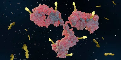
These diagnostic errors are not trivial. They can lead to inappropriate treatment choices, worsen patient outcomes, and confound clinical trials by enrolling biologically heterogeneous populations. In diseases where effective therapies are likely to depend on early intervention, misdiagnosis represents a fundamental barrier to progress.
Detecting pathology, not just protein
However, measuring alpha-synuclein reliably has proven difficult. Total protein levels in CSF are inconsistent and fail to distinguish between normal and disease-associated forms. What the field needed was a way to detect misfolded, pathogenic alpha-synuclein specifically.
That breakthrough came with seed amplification assays (SAAs), which exploit the prion-like behaviour of misfolded proteins. As Lebovitz explained, once alpha-synuclein misfolds into an abnormal shape, it can act as a “seed”, inducing normal protein to adopt the same structure. “It starts as an unstable misfolded form, but once it begins to polymerize, it becomes stable and converts other normal proteins into the same misfolded form,” he said. “This process is doubly harmful as it deprives the cell of a functional protein and generates toxic protein aggregates.”
Amprion has harnessed this knowledge and recreated it in a controlled laboratory setting. A small CSF sample is introduced into a reaction containing an excess of normal alpha-synuclein. If even minute amounts of misfolded, disease-associated protein are present, they trigger a chain reaction that converts the normal protein into the same abnormal structure.
This process is then monitored in real time using a fluorescent dye that binds specifically to the aggregated, misfolded protein. As the seeded reaction progresses, fluorescence increases, effectively amplifying a signal that would otherwise be undetectable.
This test is known as SAAmplify-αSYN, and is the first clinically available, validated test to detect pathological alpha-synuclein in CSF using seed amplification technology.
A case study in clinical translation
Across multiple studies, the test has demonstrated greater than 95 percent accuracy when compared with autopsy-confirmed diagnoses. Importantly, the test can both rule in and rule out synucleinopathy, helping distinguish Parkinson’s disease and Lewy body dementia from Alzheimer’s and other similar conditions.
Patients with amyloid and tau pathology alone progressed more slowly, while those with amyloid, tau, and alpha-synuclein declined on average three times faster.
—Russell Lebovitz, Amprion Diagnostics
One of Amprion’s earliest and most striking findings came from testing patients who had been clinically diagnosed with Alzheimer’s disease and were not thought to have any other pathology. “We found that 40 percent of these patients also showed evidence of misfolded alpha-synuclein,” said Lebovitz. “These individuals followed a very different disease course. Patients with amyloid and tau pathology alone progressed more slowly, while those with amyloid, tau, and alpha-synuclein declined on average three times faster.”
A more recent study showed that sex may further influence progression, with women experiencing dramatically accelerated decline compared with men when alpha-synuclein is present. “Now we know just in the last month or so that if you divide men and women, women may progress 10 to 20 times faster, whereas men, one and a half to two times,” said Lebovitz.

For clinicians, this biological clarity has immediate implications. Patients who appear similar in the clinic may in fact be on fundamentally different disease trajectories, with markedly different rates of progression and responses to therapy. Without molecular stratification, these differences remain hidden — often until symptoms diverge irreversibly.
Transforming clinical trials and drug development
The impact of alpha-synuclein testing may be even greater in clinical research. Many neurodegenerative trials have failed not because the drugs were ineffective, but because the wrong patients were enrolled.
If a trial for a Parkinson’s therapy includes participants who do not actually have alpha-synuclein pathology, the signal is diluted from the start. By enabling biologically precise patient selection, alpha-synuclein SAAs could improve trial design, reducing noise and accelerating decision-making.
This shift has been recognized at the highest levels. In 2024, the FDA issued a rare Letter of Support describing alpha-synuclein SAAs as a “critical drug development tool.” Fewer than 25 such letters have been issued in the past decade.
The technology is now embedded in major research efforts, including longitudinal cohorts supported by the Michael J Fox Foundation, and is being offered clinically in the US through Amprion and its commercial partner, Mayo Clinic Laboratories.
Earlier diagnosis, earlier intervention
Perhaps the most compelling promise of alpha-synuclein testing is what it enables upstream. By the time motor symptoms appear in Parkinson’s disease, an estimated 50-80 percent of dopaminergic neurons have already been lost. At that stage, disease-modifying therapies may be too late to fully prevent progression.
But alpha-synuclein pathology can be detected far earlier. “Once our CSF test becomes positive, that means the disease process has been initiated, as it’s the first thing that happens biologically,” said Lebovitz. “Clinical symptoms follow much later. In most people, it’s five to ten years before early signs like sleep disturbance or loss of smell appear, and another decade before classical motor or cognitive symptoms emerge.”
This opens a window for intervention before irreversible damage occurs.
A turning point for neurodegenerative medicine
Neurodegenerative medicine is undergoing a fundamental transition. Symptom-based diagnosis is giving way to biology-based classification, where diseases are defined by their molecular drivers rather than their clinical presentation.
Tau and amyloid have already reshaped Alzheimer’s research. Alpha-synuclein is now extending that transformation to Parkinson’s disease, dementia with Lewy bodies, and related disorders.
The challenge ahead is no longer scientific discovery, but clinical adoption: integrating molecular diagnostics into routine neurological care. If successful, the implications are profound. Fewer misdiagnoses, more targeted treatments, more successful clinical trials — and, for the first time, a realistic chance to intervene before neurodegeneration becomes irreversible.






