A glioblastoma diagnosis is tantamount to a death sentence. Most patients survive just over one year with this aggressive brain cancer. Treatment options are limited, and mortality rates have not improved for decades.
The majority of glioblastoma patients undergo surgery and then receive radiation therapy and temozolamide, the only FDA approved chemotherapy that stops the cancer’s growth. A wearable device called the Optune device extends the life of some patients with electric fields that slow cancer cell proliferation. But eventually, almost all glioblastoma patients develop resistance to both radiation and temozolamide.
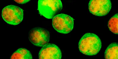
Applying a rotating magnetic field to magnetic carbon nanotubes inside a cancer cell causes the nanotubes to move.
Movie Credit: Xian Wang
Recently, an interdisciplinary team of scientists from the University of Toronto and the Hospital for Sick Children in Toronto reported an “outside-the-box” treatment for glioblastoma. Described in Science Advances, their method shows promise in a preclinical mouse model. It may provide hope for patients who once had none, but only if it can be translated into humans.
The scientists used what they call “mechanical nanosurgery” to overcome glioblastoma’s drug resistance, said Xi Huang a biologist at the University of Toronto and the Hospital for Sick Children, and coauthor of the study. He and his team collaborated with that of Sun Yu, a roboticist at the University of Toronto.
A mouse with a xenograft glioblastoma tumor undergoes focused magnetic field treatment.
Credit: Xian Wang
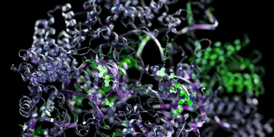
For this study, they stuffed carbon nanotubes, or CNTs, with magnetic iron nanoparticles. They also labeled the CNTs with an antibody that targets the protein CD44, which is highly expressed in many glioblastoma tumors.
To treat the mice, the researchers injected the magnetic CNTs into the brain region that contained drug-resistant glioblastoma tumors derived from human cell lines. The CNTs either entered or adhered to the surface of the cells in that region. The scientists then applied a magnetic field engineered to cause only the CNTs within the tumor region to spin. This disrupted cellular structures both inside and outside cells.
“It’s a blunt force,” said Huang. “It’s really causing mayhem in many different places.” That mayhem killed tumor cells and increased the overall survival of treated mice compared with untreated mice. Previous research showed that this strategy is an effective way to kill cancer cells, but this is the first time researchers have demonstrated it in drug-resistant glioblastoma tumors.
While the authors hope that this proof-of-concept result will translate into human patients, this may not happen anytime soon, if at all. “This is so far from the clinic right now that it's hard to know where they might stumble along the way,” said Stephen Bagley, a neuro-oncologist who specializes in glioblastoma at the University of Pennsylvania, who was not involved with the study.
As the authors point out, it is a completely novel approach — completely paradigm-shifting work. Maybe that's what is needed for glioblastoma.
- Stephen Bagley, University of Pennsylvania
Bagley said that human trials for a magnetic-CNT-based therapy might be years or even a decade away. (Huang and Yu are more optimistic about that timeline.) It is also still unclear whether this therapy might work on larger human brains with more complex human tumors. And while the majority of cells in the tumor region will be cancerous, that region will also contain some healthy brain tissue. It’s unclear how much this method will damage healthy cells.
Yu said that the CNT’s size, shape, and iron content as well as the antibodies attached to them can be finetuned to maximize their effect. But he admitted that there is a risk to healthy brain tissue. “Our goal is to be positioned to help as the last resort,” Yu said. “To deal with resistance, I think it's worth it.”
For Bagley, more immediate hope comes from other new treatments like CAR T-cell or oncolytic viral therapies that are already in Phase 1 and 2 clinical trials. But he still thinks this research shows promise. “As the authors point out, it is a completely novel approach — completely paradigm-shifting work,” Bagley said. “Maybe that's what is needed for glioblastoma.”
References
- Wang, X. et al. Mechanical Nanosurgery of Chemoresistant Glioblastoma Using Magnetically Controlled Carbon Nanotubes. Sci Adv 9, eade5321 (2023).
- Bagley, S.J. et al. Glioblastoma Clinical Trials: Current Landscape and Opportunities for Improvement. Clin Cancer Res 28, 594–602 (2022).
- Kim, D.-H. et al. Biofunctionalized magnetic-vortex microdiscs for targeted cancer-cell destruction. Nat Mater 9, 165–171 (2010).
- Liu, D. et al. Magnetoporation and magnetolysis of cancer cells via carbon nanotubes induced by rotating magnetic fields. Nano Lett 12, 5117–5121 (2012).
- Cheng, Y. et al. Rotating magnetic field induced oscillation of magnetic particles for in vivo mechanical destruction of malignant glioma. J Control Release 223, 75–84 (2016).
- Chen, M. et al. Remote control of mechanical forces via mitochondrial-targeted magnetic nanospinners for efficient cancer treatment. Small 16, e1905424 (2020).