Researchers have used light-activated silicon nanoneedles to deliver genetic material into selected individual cells, changing their behavior while leaving neighboring cells largely untouched.
The approach, developed by scientists at King's College London, could give scientists a new way to build more precise models of tissue development and disease by controlling exactly which cells receive a genetic instruction.
Published in ACS Biomaterials Science & Engineering, the study shows that porous silicon nanoneedles can concentrate laser light at the interface between the nanoneedle and a cell. This temporarily permeabilizes the cell membrane, allowing genetic cargo suspended in the surrounding medium to enter.
What could such precise control mean for disease models and drug development?
A more selective way to deliver genes
Nucleic acid delivery is already widely used to manipulate cells in research and medicine. However, most established approaches deliver genetic material to large populations of cells at once. That can be a problem when researchers want to target just a subset of cells.
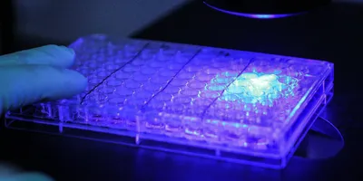
One way to achieve greater spatial precision is through optoporation, which uses precisely directed light to temporarily create openings in a cell's membrane so that genetic material can enter. By controlling where the light is directed, researchers can gain greater control over where delivery occurs.
That level of precision could eventually prove useful in several areas. In disease modeling, for example, researchers could manipulate selected cells within a larger population and study how changes in a small number of cells affect their neighbors and the wider tissue.
Greater precision could also be valuable for ex vivo cell therapies, where scientists modify cells outside the body before administering them to patients. These treatments often require researchers to first isolate and purify the cell population they want to engineer before modifying it. A technology capable of selectively manipulating particular cells within a mixed population could potentially reduce the number or complexity of those processing steps.
Tiny materials known as nanosensitizers can make optoporation more efficient by concentrating the effects of the light. Gold nanotubes and titanium dioxide nanostructures have both been used for this purpose. However, these materials can potentially damage DNA or cause other unwanted biological effects.
The King's College London team instead turned to porous silicon nanoneedles, a material with an established track record in nucleic acid delivery and a history of biocompatibility.
Tuning the needles for delivery
The researchers first had to work out how to make their tiny silicon needles as effective as possible at delivering cargo into cells.
By adjusting the laser conditions and changing the structure and surface chemistry of the nanoneedles, the team found that thermally carbonized porous silicon nanoneedles could reach up to 90 percent delivery efficiency under optimized conditions.
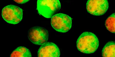
Most importantly, the researchers showed that the system could deliver functional mRNA. They introduced mRNA encoding green fluorescent protein into cells with a 72 percent delivery efficiency. Twenty-four hours later, 68 percent of the cells were producing the fluorescent protein, showing that the mRNA was successfully translated.
Changing the fate of a chosen cell
The final experiment tested whether they could program selected cells at defined locations within a larger cell population. The researchers called this “spatially resolved cell-fate engineering”.
The researchers delivered mRNA encoding SNAIL1, a transcription factor that helps drive epithelial-to-mesenchymal transition (EMT), into selected cells. EMT is an important biological process in development and wound repair, but its dysregulation has also been implicated in cancer progression and metastasis.
Following targeted delivery of SNAIL1 mRNA, the selected cells showed molecular changes consistent with an EMT-associated shift in cell state. Notably, only the cells targeted by the treatment were affected. Nearby cells remained unchanged, showing that the technique can control cell behavior with a high level of precision.
This level of control could be particularly useful for studying biological processes in which the behavior of a small number of cells has consequences for the wider tissue. Researchers could use the approach to build more precise disease models, investigate how altered cells influence their neighbors, and study how tissues develop or change over time. With further development, the same level of precision could also prove useful for selectively engineering cells for research or therapeutic applications.
However, further work is still needed before the platform can be applied to more complex biological models. The study did not include dedicated short- or long-term assessments of cell viability, and extending the approach from 2D cell cultures into organoids or other 3D tissues will present additional challenges, including light penetration and access to cells beyond the surface.