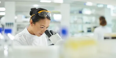
BLOOMINGTON, Ind.—Researchers at Indiana University (IU) and Harvard University have published a study revealing a new understanding of the biochemical mechanisms behind cellular division in bacteria. The study, published in the journal Science, explored the minutiae of how bacterial growth occurs and offers insights on developing drugs to combat antibiotic-resistant bacteria known as superbugs.
“This is the first time we’ve been able to observe cell division as a dynamic process—that is, a process occurring over time,” said Erkin Kuru, a former IU Ph.D. student. “This wasn’t possible before since we lacked the tools to see it.”
The discoveries were made using IU’s groundbreaking method for coloring bacterial cell walls developed in 2012. The technology allows researchers to apply different colored dyes, called fluorescent D-amino acids, or FDAAs, to a cell at different times, resulting in a multicolored canvas that illustrates the timeline and process of bacterial cell wall production. When announcing the new method, IU researchers were aware of the potential to develop new antibiotics based on the added understanding, and the recent paper demonstrates its promise.
“This is the first study to ‘connect the dots’ between each part of the cell involved in bacterial cellular division,” said Yves Brun, the Clyde Culbertson Professor of Biology in the IU Bloomington College of Arts and Sciences’ Department of Biology, who is an author on the study. “We’ve finally closed the circle on this mechanism and opened the door to more precise methods in the fight against antibiotic-resistant bacteria.”
Antibiotic-resistant bacteria are a global concern, with one in four hospital-acquired infections in long-term U.S. patients caused by six major superbugs. Bacteria can carry genes that allow them to survive exposure to our existing stable of antibiotics, meaning that these infections are harder to treat, although they are not necessarily more severe or infectious. Adding to the peril of these bacteria is that the antibiotic resistance gene can be passed between bacteria, permitting creation of a bacteria that can be resistant to many antibiotics, known as a superbug.
The IU/Harvard discovery utilized the FDAA coloring to visualize cell division, illuminating new understanding in the building of cell walls. Parts of the cell that drive bacterial division include cell wall-making enzymes called penicillin-binding proteins, or PBPs, and cytoskeletal proteins, called FtsA and FtsZ, which form skeleton-like fibers inside cells to direct construction of the wall. These elements must coordinate to build a cell wall in the middle of the cell to ensure the material inside doesn’t escape after it splits in half. This study is the first to show exactly how they coordinate. Essentially, FtsZ acts as a “foreman” that directs the movement of PBP “workers” as they construct a cell wall.
“The application of different colors of these dyes during the cell wall construction process revealed a ‘bull’s-eye pattern,’ indicating the circular wall is built from the outer edge of the cell inward to the center,” said Michael VanNieuwenhze, a professor in the IU Bloomington College of Arts and Sciences’ Department of Chemistry, and co-author on the study.
The study also solves another mystery: How do FtsZ molecules build the wall? The researchers found that FtsZ—which is arrayed in a biochemical chain called a filament—constantly loses a molecule at one end and gains a molecule at the other end, resulting in a circular motion around the cell’s edge described as “treadmilling.”
Through a better understanding of how superbugs might multiply, researchers and drug developers may be able to arrest the process with a powerful impact on global health.
“If you understand how an engine works, you can shut it down by removing a single part,” Brun said. “You no longer need to throw a hammer into the works to destroy it.”