Cancer cells are the mothers of genetic invention. Desperately shredding, rewiring, and reconfiguring their own genomes, tumor cells evade immune detection, scramble for blood, oxygen, and nutrients, and evolve in a thousand directions at once in a twisted Darwinian struggle against our own bodies.
The seemingly endless creativity and diversity of cancer can make it a difficult target for medicines. Made of fundamentally human cells, cancer can be difficult to target in the clinic without also killing healthy cells. Cancer cells camouflage themselves as normal cells to bypass immune surveillance. The similarity of cancer cells to the cells of the body they grow in was so ingrained in the minds of researchers that for years — as late as 1976 — many thought that the immune system couldn’t react to tumors at all (1). But the genetic inventiveness of cancer leaves researchers and doctors with a unique opportunity, a potentially never-before-seen, never-to-be-seen-again target: the neoantigen.
Neoantigens are proteins and other peptides expressed by cancer cells from mutated genes created during the transformation of normal, healthy cells into cancer cells. These proteins are not expressed by normal cells. They may not even be shared between patients who have the same kind of cancer. Because of that, neoantigens present a sterling opportunity to precisely target cancer with a small molecule drug or a vaccine with minimal side effects.
The idea that cancer cells are mutants dates to 1902, when Theodor Boveri, an early cell biologist at the University of Würzburg, postulated that cancer cells arise when healthy cells scramble their chromosomes, an electrifying idea at a time when DNA wasn’t yet known as the carrier of genetic information. From there, however, it took a disquietingly long time to paint a fuller picture.
In 1953, EJ Foley, a cancer biologist at Schering Corporation (now part of Merck), showed that immune cells specifically recognize tumors (2). Whether that meant that immune systems actually fight against cancer still wasn’t clear though. It took another 30 years to show that immune systems have full-blown responses to tumor cells. The cancer geneticists Aline Van Pel and Thierry Boon at the Ludwig Institute for Cancer Research created a cancer cell line that not only stimulated a mouse’s immune system, but also raised an immune response that was specific to that tumor (3). A new paradigm emerged: immune systems are capable of responding to cancer, but because of immune evasion techniques employed by cancer cells, they are often deprived of novel epitopes to target.
In the mid-1990s, a joint German and American research group led by the immunologist Thomas Wölfel from Johannes Gutenberg University separated and sequenced DNA samples from a melanoma tumor. Among the pools of sequences, they identified a single-base change in a 303-base-long sequence that turned out to encode the gene for cyclin dependent kinase 4 (CDK4), a cell cycle regulatory protein now known as one of the most commonly mutated proteins in human cancers. The finding was so painstakingly won and so novel for the time that the raw sequencing gels showing the mutation appeared front and center in a characteristically dense and cramped Science paper (4).
A few years later in 2004, a group led by immunobiologist Paul Robbins at the National Institutes of Health observed that the near complete regression of a melanoma patient’s tumors resulted from T cells recognizing a neoantigen (5). The tediousness and laboriousness of dealing with genomes in the age before next generation sequencing made picking novel protein-coding sequences out of the muck of a tumor’s genome difficult, so research on neoantigens quieted for about 10 to 15 years after that. But, with the introduction and increasing affordability of next generation sequencing in the 2000s and 2010s, experimental data began piling up showing the potency of neoantigens for eliminating tumors.
A missed opportunity
Neoantigens offer a new array of bullseyes for therapeutics, but in the 21st century, the field is just catching up to the idea that oncogenic mutations in the genome produce protein mutations that T cells can be trained to attack. The therapeutic potential of neoantigens was obscured not only by history, but also by the models scientists use to study cancer in the lab.
“The genomic landscape of somatic mutations [in common laboratory models] is extremely quiet. These models lack potential neoantigens. So, in the cases of lung cancer, melanoma, colon cancer, they fail to recapitulate the mutational heterogeneity and diversity of their human counterparts,” said Peter Westcott, a cancer biologist at Cold Spring Harbor Laboratory. “There’s copy number instability, etc., but in regard to neoantigens, very little.”
For mutations to progress into full blown cancer, they usually occur in five to 10 “driver” genes involved in cell growth or genome maintenance, with hundreds or thousands of “passenger” mutations that don’t cause malignancy occurring alongside. Ninety percent of neoantigens arise from passenger mutations (6). Since artificially induced tumors lacked this heterogeneity, for years the idea of picking out cancer-specific or even patient-specific mutations became an afterthought.
To make laboratory created and patient derived mutations match, oncologists are letting laboratory cells mutate in a way that more realistically mimics tumor development in the body. “One of the things I'm doing is targeting DNA repair pathways to generate mutational instability, and as these tumors continue to accrue mutations, see how that elicits T cell responses and how the tumors evade that,” said Westcott.
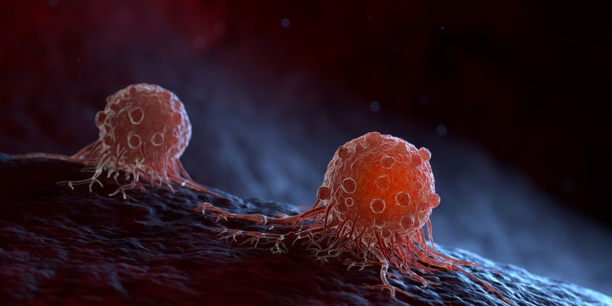
As cells become cancerous they express mutated genes created from their transformation from healthy cells to cancerous ones. These mutated genes are translated into new proteins called neoantigens.
Credit: iStock/iLexx
Mutations may produce cancer cells, but full blown disease doesn’t arise unless the body cannot repair those mutations. It’s not surprising then that damage to DNA repair pathways is one of the most common drivers of mutations across all tumor types, speeding what initially might be a handful of mutated cells into tumors and metastases (7). Patients with mutations in DNA repair pathways are at some of the highest risks of developing hard-to-treat malignancies. These patients harbor “microsatellite instable” mutations, which can predispose them to specific kinds of cancer (8).
While cancers with large numbers of mutations can be deadly and aggressive, their mutations can also be an Achilles heel. Despite displaying a large number of potential neoantigens for T cells to recognize, these tumors often evade immune surveillance by also displaying immune checkpoint peptides. Immune cells display a protein on their surface, like programmed-cell death protein 1 (PD-1) and if a cell displays a corresponding ligand that the protein can bind, the immune cell will not attack. Only healthy cells produced by the body should display these peptides, and if they do, immune cells will pass over them. Tumors can produce checkpoint protein ligands en masse, effectively hiding themselves from the immune system. The success of immunotherapies such as checkpoint inhibitors, which block tumor cells’ abilities to evade immune surveillance, directly correlates with mutational burden, DNA-repair insufficiency, and neoantigen production (9,10).
What’s real?
DNA-repair deficient cancers should generate reams of novel sequences, but just because a tumor generates lots of mutations doesn’t mean that it produces a neoantigen. Not every protein sequence can reliably raise an immune response, so the question becomes whether neoantigens are a widespread oncological phenomenon or are just a feature of particularly mutation-laden microsatellite unstable cancers.
As a tumor grows and is attacked by the body, the composition of the tumor evolves over time, and the cells and antigens that the immune system sees change. Cancer cells that survive a round of immune system attacks can discard a previous generation of neoantigens, so a tumor sample taken at one time point might be out of date down the road.
“All those mutations are shared in essentially every single tumor cell growing out in that tumor, so that can greatly change the landscape of the immune response against those new antigens,” said Westcott.
All those mutations are shared in essentially every single tumor cell growing out in that tumor, so that can greatly change the landscape of the immune response against those new antigens.
- Peter Westcott, Cold Spring Harbor Laboratory
Charting a path for neoantigen discovery requires charting the evolution of tumors in a body, according to Alex Jaeger, a cancer biologist at the Moffitt Cancer Center. That starts with better models.
“It's very difficult to go into a patient and ask whether or not a particular treatment is directly influencing what peptides their cancer cells are presenting. But in the mouse, we can actually do those experiments. We can evaluate a panel of drugs and directly ask how they're modulating the tumor microenvironment to change the antigenicity of cancer cells in vivo,” said Jaeger.
“When we initiate tumor formation, we can actually evaluate what peptides are being presented through tumor evolution,” said Jaeger. “In the earliest stages of transformation prior to invasive disease, are there different peptides that are being presented on those cells that could be targets for early intervention? Or prophylactic vaccines that could intercept cancer before it actually progresses to a more invasive disease?”
Individual therapies for individual cells
A prophylactic vaccine using a neoantigen is a tantalizing idea. Immunizing patients with proteins or mRNA encoding a cancer-specific peptide could stop a metastasis before it even starts. But knowing what neoantigens to immunize against is still an open question.
In 2008, a group led by the geneticist Richard Wilson at Washington University in St. Louis produced the first full cancer genome for acute myeloid leukemia (11). This established a first full cross-section of alterations in protein-coding genes, adding new momentum to the field. A scant four years later in 2012, a group of immunologists led by Robert Schreiber also at Washington University in St. Louis published the first comprehensive profile of neoantigens in a tumor (12).
A number of neoantigen cancer vaccines developed in the 2000s and 2010s have already advanced to clinical trials. One targeted a protein called gp100 that is frequently altered in melanoma (14). Another targeted NY-ESO-1 protein in a variety of tumor types. Yet another was tested in children with neuroblastomas and sarcomas (15). However, no vaccine targeting a neoantigen has succeeded in a phase II clinical trial.
“All these vaccine trials failed,” said Matthew Gubin, an immunologist at the MD Anderson Cancer Center. “One of the reasons I think is that they vaccinated, but they weren't combined with any kind of immune checkpoint. And so you're getting the ON signal, but the T cells are getting shut off easily.” Even with the glaring neon sign reading “attack this tumor” that a vaccine provides, cancer cells can still evade an immune response and grow.
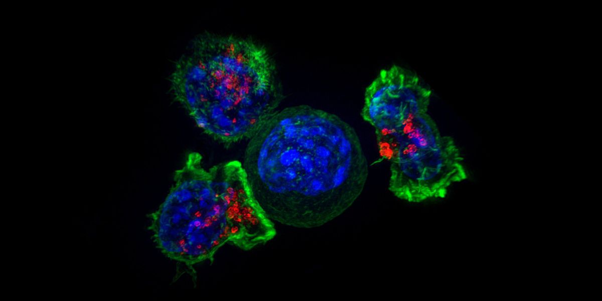
Killer T cells surround and attack a cancer cell.
Credit: NICHD/J. Lippincott-Schwartz
Even when combined with checkpoint inhibitors, there are still problems. Sequencing the genome of a tumor, for instance, is an act of prediction for an oncologist. Just because a gene is mutated does not mean that the protein that gene encodes is expressed or available as an antigen. How can scientists catch a never-before-seen peptide red-handed?
This is an active field of research with no clear answers, but some researchers are turning to artificial intelligence (AI) programs to analyze and predict immunogenic neoantigens to produce vaccines. In 2022, one group at the Copenhagen University Hospital led by the oncologist Inge Marie Svane created a vaccine consisting of five to 10 different neoantigen peptides produced in people with melanoma. The vaccines were manufactured within 60 days of the tumor biopsy (16). Although this was a small trial with five participants, that vaccine successfully raised a T cell response without adverse effects.
With decades of research in hand, the ability to sequence DNA, RNA, and proteins and the details of antigen presentation, tumor evolution, and gene repair coming into somewhat clearer view, the lofty goal is for cancer treatment to look very different in 25 to 50 years from how it looks today.
“Patients would come in. They have cancer, a piece of which you need less and less material of now to do sequencing. You get a biopsy,” said Gubin. From there, a laboratory can look for a host of known mutations, insertions, deletions, and frameshifts. “You'll come up with the list,” said Gubin. An oncologist could put together a plan with a combination therapy with checkpoint blockade inhibitors, chemotherapy, and a custom vaccine. And while it can be difficult to synthesize proteins on an individual basis to produce a traditional protein-based vaccine, the age of mRNA vaccines has made this hurdle less concerning.
Prophylactic cancer vaccines for at risk patients may even be in the offing. If a patient has known cancer-causing mutations, safe is better than sorry. “We might as well vaccinate because they have lung cancer or pancreatic that runs in their family and tend to have these types of mutations,” said Gubin. “So that could be a possibility if that makes sense.”
References
- Hewitt, H. B., Blake, E. R. & Walder, A. S. A critique of the evidence for active host defence against cancer, based on personal studies of 27 murine tumours of spontaneous origin. Br J Cancer 33, 241–259 (1976).
- Foley, E. J. Antigenic properties of methylcholanthrene-induced tumors in mice of the strain of origin. Cancer Res 13, 835–837 (1953).
- Van Pel, A. & Boon, T. Protection against a nonimmunogenic mouse leukemia by an immunogenic variant obtained by mutagenesis. Proc Nat Acad Sci USA 79, 4718–4722 (1982).
- Wölfel, T. et al. A p16 INK4a -Insensitive CDK4 Mutant Targeted by Cytolytic T Lymphocytes in a Human Melanoma. Science 269, 1281–1284 (1995).
- Huang, J. et al. T Cells Associated with Tumor Regression Recognize Frameshifted Products of the CDKN2A Tumor Suppressor Gene Locus and a Mutated HLA Class I Gene Product. The Journal of Immunology 172, 6057–6064 (2004).
- Kantoff, P. W. et al. Sipuleucel-T Immunotherapy for Castration-Resistant Prostate Cancer. N Engl J Med 363, 411–422 (2010).
- Kiwerska, K. & Szyfter, K. DNA repair in cancer initiation, progression, and therapy—a double-edged sword. J Appl Genetics 60, 329–334 (2019).
- Lynch, H. et al. Review of the Lynch syndrome: history, molecular genetics, screening, differential diagnosis, and medicolegal ramifications. Clinical Genetics 76, 1–18 (2009).
- Le, D. T. et al. Mismatch repair deficiency predicts response of solid tumors to PD-1 blockade. Science 357, 409–413 (2017).
- Yarchoan, M., Hopkins, A. & Jaffee, E. M. Tumor Mutational Burden and Response Rate to PD-1 Inhibition. N Engl J Med 377, 2500–2501 (2017).
- Ley, T. J. et al. DNA sequencing of a cytogenetically normal acute myeloid leukaemia genome. Nature 456, 66–72 (2008).
- Matsushita, H. et al. Cancer exome analysis reveals a T-cell-dependent mechanism of cancer immunoediting. Nature 482, 400–404 (2012).
- Castle, J. C. et al. Exploiting the Mutanome for Tumor Vaccination. Cancer Research 72, 1081–1091 (2012).
- Rosenberg, S. A. et al. Immunologic and therapeutic evaluation of a synthetic peptide vaccine for the treatment of patients with metastatic melanoma. Nat Med 4, 321–327 (1998).
- Krishnadas, D. K. et al. A phase I trial combining decitabine/dendritic cell vaccine targeting MAGE-A1, MAGE-A3 and NY-ESO-1 for children with relapsed or therapy-refractory neuroblastoma and sarcoma. Cancer Immunol Immunother 64, 1251–1260 (2015).
- Mørk, S. K. et al. Personalized therapy with peptide-based neoantigen vaccine (EVX-01) including a novel adjuvant, CAF®09b, in patients with metastatic melanoma. OncoImmunology 11, 2023255 (2022).