Genetic screens can give researchers all sorts of insights into how a particular small molecule or drug candidate might act in a given disease. While these kinds of screens work well when scientists are working with a cell type that they can easily culture and expand, their task becomes much harder if they want to perform the same experiments in rare patient cells or cells that are more difficult to culture. Also, certain cell types may require specific reagents to sustain them, increasing the cost of the screen.
Alison Hirukawa and her team at DropGenie, of which she is the co-founder and Chief Executive Officer, have developed a tool to enable high-throughput gene-editing screens in these valuable cells. Their technology allows scientists to perform experiments in more translationally relevant cell types and use fewer cells, decreasing costs.
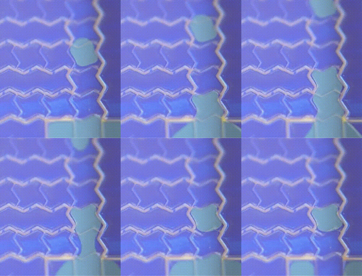
Using digital microfluidics, the team at DropGenie can perform gene editing screens with small numbers of cells.
Credit: DropGenie
“We can enable any [gene-editing] payload into any cell type to enable any type of cell engineering,” said Hirukawa. “Every edit is happening in its own well, or cubicle, or — in our case — droplet, and that really enables you to start layering the multiomic data sets that you pull out.”
The researchers create these individual droplets using digital microfluidics, which, as Hirukawa explained, “actually should be called ‘electrostatic forces microfluidics,’ but that doesn't sound very interesting.” Basically, the team designed a plate that has tiny clusters of electrodes with a hydrophobic coating. When the researchers turn the tiny areas of electrodes on or off, they create a voltage, which can create droplets that are one microliter in size. The team can then use that voltage to manipulate the position of the droplet.

Rather than using a virus to deliver the gene-editing machinery to cells in their screens, the DropGenie researchers take advantage of their digital microfluidic electrical environment to deliver their gene-editing payloads via a gentler electroporation protocol. Their technique successfully edited primary human T cells (1). In a recent preprint, the team made a head-to-head comparison with their device and two other commercially available electroporation devices (2). They found that their system, using fewer T cells and a lower amount of the genetic payload, led to less genetic dysregulation in the edited T cells.
We can enable any [gene-editing] payload into any cell type to enable any type of cell engineering.
– Alison Hirukawa, DropGenie
Hirukawa and her team recently tackled another, even more rare, type of immune cell: regulatory T cells (Tregs). “There's no good immortalized cell model of these, so people really want to understand the biology of Treg cells,” she said. “We were able to hone into a protocol set that gave us over 85 percent knockout of our target locus in these cells. And we did that with about a million cells total. [With] other platforms, you need a million cells for one edit.”
The team is now implementing knock-in edits into their system; these are more complex edits to make than just knocking a gene out. They’re also expanding into other cell types like neural cells and multicellular structures such as organoids and other 3D cell models. They have plans to roll out an even higher throughput platform later this year.
“It's so exciting and such a privilege to enable all these drug hunters and folks that are working on these really, really difficult human health questions,” said Hirukawa. “Tools are really only going to become more and more powerful. I think this is a very exciting time to be in a tools company.”
References
- Little, S.R. et al. A Tri-Droplet Liquid Structure for Highly Efficient Intracellular Delivery in Primary Mammalian Cells Using Digital Microfluidics. Adv Mater Technol 8, 2300719 (2023).
- Little, S.R. et al. A Digital Microfluidic Platform for the Microscale Production of Functional Immune Cell Therapies. Preprint at bioRxiv (2024).