European hematology laboratories are asking the same question: as next-generation sequencing (NGS) panels expand genomic coverage, at what point does fluorescence in situ hybridization (FISH) become redundant? Clinical laboratories managing chronic lymphocytic leukemia (CLL), multiple myeloma (MM), and hematological malignancies broadly are navigating a simultaneous technology and regulatory choice, as the European Union's (EU) In Vitro Diagnostic Regulation (IVDR) forces decisions about which FISH probes to retain and re-certify before compliance deadlines expire. The evidence on FISH's continued clinical necessity, however, is clearer than the market dynamics suggest.
OGT announced IVDR certification for four additional CytoCell fluorescence in situ hybridization probes, bringing its total of IVDR-certified CytoCell probes to 22. The newly certified probes expand hematological malignancy coverage under IVDR with markers relevant to non-Hodgkin lymphoma, acute lymphoblastic leukemia (ALL), CLL, and MM; all four were previously available under the older IVD Directive (IVDD) framework and have been recertified as part of OGT's program to transition its full CytoCell IVDD portfolio ahead of the Class C deadline of December 31, 2028.
IGH translocations and ATM deletion: why FISH holds
Two biological features make these markers structurally resistant to NGS displacement: the heterogeneous breakpoint architecture of IGH rearrangements, and the turnaround demands of CLL management. The immunoglobulin heavy chain (IGH) locus on chromosome 14q32 is the most recurrent site of pathogenic translocation in B-cell malignancies. Its translocations involve breakpoints distributed across large genomic intervals with no fixed junction, requiring careful bioinformatic handling in NGS that reduces sensitivity compared with locus-specific FISH probes on plasma cell-enriched bone marrow samples.
In CLL, deletion of the ATM (ATM serine/threonine kinase) gene at 11q22.3 cooperates with TP53 loss to impair the DNA damage checkpoint, conferring poor prognosis and influencing the choice between chemoimmunotherapy and targeted agents. FISH returns a result within hours; NGS workflows in CLL typically take one to two weeks. Speed is a clinical requirement in CLL, not a preference.
In MM, IGH translocations with partner loci, including FGFR3 (fibroblast growth factor receptor 3) at 4p16.3, which drives the t(4;14) rearrangement, and MYEOV (myeloma overexpressed) at 11q13.3, are required by International Myeloma Working Group (IMWG) risk models at diagnosis. A 2025 Blood Cancer Journal guidelines paper from the Cancer Genomics Consortium Plasma Cell Neoplasm Working Group, led by Xinyan Lu and co-senior-authored by Shaji Kumar at the Mayo Clinic's Division of Hematology and Medical Oncology, confirmed that FISH remains the gold-standard clinical assay for detecting genetic abnormalities in MM, noting that panel design and reporting practices vary among laboratories. A companion 2025 HemaSphere consensus from the European Myeloma Network (EMN), led by Niccolò Bolli of the University of Milan, recommended NGS as a complement rather than a replacement for FISH, citing cost, accessibility, and workflow integration as barriers to substitution.
Why plasma cell biology favors FISH
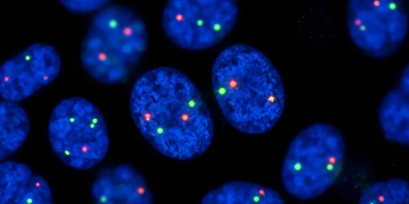
Plasma cells in MM do not enter mitosis reliably in vitro, making FISH on interphase nuclei the primary route to cytogenetic characterization from bone marrow aspirates. FISH also performs well at low tumor cell fractions when plasma cell enrichment is applied, and its signal is interpretable at the single-cell level, enabling clone size estimation that population-averaged sequencing depth cannot replicate. Interphase capability and single-cell resolution keep FISH as the required first tier for IGH and ATM characterization regardless of NGS platform availability.
Four probes: specifications and clinical scope
Each of the four newly certified probes addresses a distinct diagnostic gap, covering IGH screening, ATM deletion, and two specific IGH partner translocations with therapeutic implications:
- CytoCell ATM Deletion FISH Probe (CE-LPH 011): Detects deletion at 11q22.3 in CLL; includes a centromeric chromosome 11 control to distinguish deletion from monosomy.
- CytoCell IGH Plus Breakapart FISH Probe (CE-LPH 070): Detects any IGH (14q32.3) rearrangement regardless of partner locus, supporting broad screening in CLL, lymphoma, and ALL.
- CytoCell IGH/FGFR3 Plus Translocation, Dual Fusion FISH Probe (CE-LPH 074): Detects the t(4;14)(p16.3;q32.3) rearrangement in MM, classified as an adverse-risk cytogenetic feature under IMWG and International Myeloma Society (IMS) 2025 risk stratification criteria.
- CytoCell IGH/MYEOV Plus Translocation, Dual Fusion FISH Probe (CE-LPH 078): Detects the t(11;14)(q13;q32) translocation in MM and lymphoma; t(11;14) carries standard-risk designation in myeloma and is of growing predictive interest for venetoclax sensitivity, driven by BCL2 upregulation at the rearranged locus.
All four were previously available under IVDD certification. Existing CytoCell users switching to the IVDR-certified equivalents do not require laboratory revalidation, removing a significant barrier for labs managing multiple simultaneous test transitions.
IVDR Class C deadline: what it means for FISH probe selection
EU laboratories must switch to an IVDR-certified probe once one is commercially available for a given intended purpose, making the December 31, 2028 Class C deadline a practical obligation for hematological malignancy FISH testing. IVDR replaced the EU's prior IVD Directive and became applicable in May 2022, introducing mandatory independent notified-body review for a larger proportion of diagnostic devices than the directive had required. CytoCell FISH probes for hematological malignancies are classified as Class C devices under the regulation.
For FISH probe manufacturers, the deadline forces a choice between full portfolio recertification and selective retirement of legacy probes. OGT reports committing to the former. The company states it was the first FISH probe manufacturer to achieve IVDR certification, having begun discussions with notified body BSI in 2019.
Tori Anthony-Dubernet, Director of European Sales and Support at OGT, who trained as a registered clinical cytogeneticist before moving into the commercial sector, described the transition challenge. "Clinical laboratories are balancing increasing regulatory requirements with the need to maintain efficient, high-quality testing workflows," Anthony-Dubernet said. "We're very happy to be expanding our IVDR-certified CytoCell portfolio with these important FISH probes and to help laboratories transition to IVDR with confidence."
The table below shows selected probes from the CytoCell IVDR portfolio, spanning the four newly certified additions and previously certified probes across CLL, MM, and leukemia indications:
| Probe | Supported malignancy | Chromosome region | IVDR status |
|---|---|---|---|
| ATM Deletion (CE-LPH 011) | CLL: del(11q22.3) | 11q22.3 | Newly certified |
| IGH Plus Breakapart (CE-LPH 070) | CLL, lymphoma, ALL: IGH screening | 14q32.3 | Newly certified |
| IGH/FGFR3 Plus, Dual Fusion (CE-LPH 074) | MM: t(4;14)(p16.3;q32.3) | 14q32.3 / 4p16.3 | Newly certified |
| IGH/MYEOV Plus, Dual Fusion (CE-LPH 078) | MM, lymphoma: t(11;14)(q13;q32) | 14q32.3 / 11q13.3 | Newly certified |
| MLL (KMT2A) Breakapart (CE-LPH 013) | ALL, AML, MDS | 11q23.3 | Previously certified |
| P53 (TP53)/ATM Combination Deletion (CE-LPH 052) | CLL: del(17p13) and del(11q22.3) | 17p13 / 11q22.3 | Previously certified |
| IGH/MAF Plus v2, Dual Fusion (CE-LPH 108) | MM: t(14;16) | 14q32.3 / 16q23 | Previously certified |
| CKS1B/CDKN2C (P18) Amplification/Deletion (CE-LPH 039) | MM: 1q21 amp / 1p32 del | 1q21-q22 / 1p32.3 | Previously certified |
FISH and NGS in hematological malignancy: the case for parallel use
FISH and NGS detect largely non-overlapping sets of clinically relevant variants and function as sequential or parallel layers of a diagnostic architecture, not competing methods. At the single-cell level, targeted structural variants in non-dividing cells are exactly where FISH excels, without requiring the bioinformatic infrastructure clinical NGS demands. NGS provides mutational profiling, higher-resolution copy-number analysis, and detection of variants no probe covers, extending rather than duplicating FISH's reach.
A 2025 research letter in the British Journal of Haematology, comparing FISH to the Myeloma Genome Project targeted sequencing panel (MGPP) in 50 newly diagnosed MM patients, showed that sequencing reclassified 22 percent of standard-risk patients to high-risk when reviewed by a blinded 15-clinician multidisciplinary team. The reclassification was driven primarily by bi-allelic TP53 inactivation and 1p32 deletion that FISH had missed, while FISH remained the initial risk-assignment tool for all patients.
When FISH signals need confirmation
Atypical signal patterns in complex rearrangements can produce misleading results, and confirmatory sequencing is warranted when patterns are unusual or precise partner identification affects treatment. A 2024 Clinical Pathology study by Prasad Koduru and colleagues at UT Southwestern Medical Center showed that atypical FISH signals led to misdiagnosis in three hematological malignancy cases, each corrected by NGS or RNA sequencing. The findings reinforce the value of FISH as a rapid first-tier screen and sequencing as a confirmatory layer, rather than framing the two as alternatives.
FISH remains essential for hematological malignancy risk stratification as IVDR reshapes the probe market
As the December 31, 2028 IVDR deadline concentrates the probe market toward certified products, the clinical evidence for FISH in hematological malignancy risk stratification has not weakened. Two 2025 consensus papers, from the EMN and the Cancer Genomics Consortium, explicitly position FISH as the required first-tier assay for structural variant detection. OGT's program to certify its full hematological malignancy catalog under IVDR, spanning probes for ALL, acute myeloid leukemia (AML), acute promyelocytic leukemia (APL), CLL, chronic myeloid leukemia (CML), lymphoma, myelodysplastic syndrome (MDS), MM, and myeloproliferative neoplasms (MPNs), offers laboratories a route to IVDR compliance without replacing the probes their workflows already depend on.
This article is based on a press release issued by OGT and was produced under Drug Discovery News' AI Editorial Guidelines.