inner ear

An MRI image of a mouse head showing the brain in gray, the cerebrospinal fluid in blue, and the fluid of the inner ear in green.
| 5 min read
A mouse cochlea is seen with hair cells colored green and neurons stained in red, twisting against a black backdrop.
| 9 min read
Subscribe to Newsletter

Subscribe to our eNewsletters

Stay connected with all of the latest from Drug Discovery News.

Subscribe

Sponsored

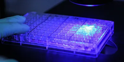
Gloved hand holding a multiwell cell culture plate under a fluorescence microscope for spheroid analysis.
Using confocal live-cell imaging to characterize immune-mediated cytotoxicity over time.
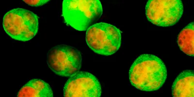
Fluorescence microscopy image of cells displaying green and red fluorescent signals.
Using confocal live-cell imaging to assess treatment effects in spheroids, organoids, and co-cultures.
3D illustration of Y-shaped antibody molecules floating against a soft blue background, with a detailed antibody structure in the foreground.
From molecular developability to functional activity, integrated characterization strategies help inform lead selection and optimization in biologics discovery.