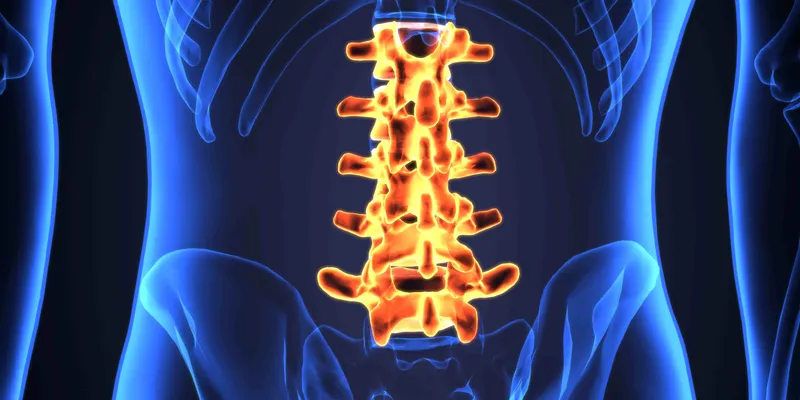
When a thin ligament in the spine thickens and bulges into the central canal, it compresses spinal nerves and leads to pain, numbness, and mobility issues. That’s what happens in lumbar spinal stenosis (LSS), which affects an estimated 103 million people worldwide.
Disease-modifying therapies for LSS remain limited to surgical intervention, primarily because the underlying biology has long been a mystery. Hoping to change that, researchers at Weill Cornell Medicine and the Hospital for Special Surgery (HSS) set out to break down why the ligament undergoes hypertrophy. Reasoning that abnormal growth begins with activated, tissue-resident stem cells producing excess ligament cells, the team sought to identify the stem cells responsible for forming tendons and ligaments.
In a study published in Cell, the team successfully identified these universal stem cells and confirmed that their hyperactivity in the lower spine drives LSS. As Matthew Greenblatt, Associate Professor of Pathology and Laboratory Medicine at Weill Cornell Medicine and senior co-author of the paper, explained to DDN, “There has been almost no mechanistic study of this disease.” That has made it nearly impossible to design targeted nonsurgical therapies. The new research paves the way for new clinical approaches, including repurposing existing blood pressure medications to target the condition at its root.
Identifying tendon and ligament stem cells in humans and mice
Because tendons and ligaments contain similar, hard-to-distinguish fibroblast-like cells, identifying true stem cells has been difficult. While earlier studies suggested potential candidates, none proved that a single population could self-renew and generate all tendon and ligament cell types.
To isolate stem cells — often referred to as the body’s master cells — researchers digested tissue into single cells and used flow cytometry alongside cell sorting to map distinct marker profiles. By transplanting different cell subsets to observe who makes who, in a sense, the team pinpointed the exact population at the top of the cellular hierarchy.
There has been almost no mechanistic study of this disease.
—Matthew Greenblatt, Weill Cornell Medicine
To confirm their findings in vivo, the team used lineage tracing and a special barcoding system to track mutations during cell division and map cell fates over time. Greenblatt said that these experiments provided “key information that really gave us confidence that we'd identified the stem cell.”
In mice, the team discovered these stem cells reside in a specialized area within tendon and ligament tissue that serves as a reservoir for tissue renewal. Using this knowledge, they identified a similar population of stem cells in human ligament samples obtained during surgery. Lingling Hu, a postdoctoral fellow and study co-author, confirmed that these human stem cells could also self-renew and generate new ligament cells, highlighting their clinical significance.
Mechanical and molecular triggers of LSS
Drawing on clinical insights from Sravisht Iyer, Associate Professor of Orthopedics at Weill Cornell Medicine and spine surgeon at HSS and study co-author, the researchers recognized that spinal hypermobility acts as a primary risk factor for LSS. Greenblatt explained, “When there is hypermobility of a spinal segment, that hypermobility is a risk factor, a substrate for developing that spinal stenosis.”
Hypermobility triggers a biological cascade that induces calcium flux in stem cells and activates calcium-responsive transcription factors, driving tissue overgrowth. Along those lines, the researchers were able to trigger tissue overgrowth by increasing calcium signaling in stem cells from healthy ligaments using genetic manipulation. This finding demonstrated that this molecular pathway alone is sufficient to cause the abnormal growth seen in LSS.
When comparing ligament tissue from patients with LSS to those with herniated discs, researchers found a significantly higher concentration of stem cells in LSS tissue. Furthermore, when transplanted into mice, these stenosis-derived stem cells produced far more output cells than those from healthy tissue — confirming their direct role in driving disease pathogenesis.
Therapeutic applications
With this new mechanistic information in hand, the researchers then showed that repurposing blood pressure drugs to block calcium signaling prevented ligament hypertrophy in animal models of spinal stenosis. While clinical trials are needed, these hypertension drugs could offer a promising nonsurgical treatment for LSS.
Because this universal stem cell gives rise to all tendon and ligament tissue, Greenblatt emphasized that its clinical potential reaches far beyond LSS.
“This cell should be at the heart of probably all or most tendon and ligament disorders, of which spinal stenosis is only one,” he noted, pointing to a future where these insights drive novel treatments for inflammatory tendinopathies, sports injuries, and chronic degenerative conditions.