MADISON, Wis.—Stem cells have, of course, offered (and continue to offer) much promise and potential over the years—as well as some practical uses already in therapeutics—but it is that potential that is often the rub in life-sciences research and pharma/biotech. The end goal, whether disease modeling or therapeutics or something else, is visible, but the path to get there turns out to be quite a bit less easily navigable than researchers might have hoped.
One important area in which researchers have as yet been unsuccessful is in the effort to produce cells that will ultimately create functional arteries. The ability to give rise to healthy arteries from stem cells would be a huge boost in fighting cardiovascular disease.
As Brian Mattmiller wrote recently for the Morgridge Institute for Research website, new techniques developed at a Morgridge Institute for Research and the University of Wisconsin-Madison (UW-Madison) have produced, for the first time, functional arterial cells at both the quality and scale to be relevant for disease modeling and clinical application.
Scientists in the lab of James Thomson, one of the stem cell research world’s pioneers, described in the July 10 issue of the Proceedings of the National Academy of Sciences methods for generating and characterizing arterial endothelial cells, which initiate artery development. These cells are said to exhibit many of the specific functions required by the body.
Further, Mattmiller writes, “these cells contributed both to new artery formation and improved survival rate of mice used in a model for myocardial infarction. Mice treated with this cell line had an 83-percent survival rate, compared to 33 percent for controls.”
“The cardiovascular diseases that kill people mostly affect the arteries, and no one has been able to make those kinds of cells efficiently before,” says Jue Zhang, a Morgridge assistant scientist and lead author. “The key finding here is a way to make arterial endothelial cells more functional and clinically useful.”
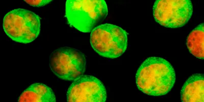
The Thomson lab has made arterial engineering one of its top research priorities, as noted on the Morgridge website, because the key challenge in this effort has not been creating the cells. Generating endothelial cells is relatively easy, but those cells lack actual arterial properties, being too “generic” and thus having very little clinical value. So, how did the team overcome this challenge and create cells that have many of the actual arterial cell functions? They applied two key technologies to the project, using single-cell RNA sequencing to identify the signaling pathways critical for arterial endothelial cell differentiation—they found about 40 genes of “optimal relevance” doing this—and then using CRISPR-Cas9 gene-editing technology that allowed them to create reporter cell lines to monitor arterial differentiation in real time.
“With this technology, you can test the function of these candidate genes and measure what percentage of cells are generating into our target arterial cells,” says Zhang.
The research group developed a protocol around what seem to be the five key growth factors most strongly contributing to arterial cell development, also identifying some common growth factors used in stem cell science (such as insulin) that, as wrote Mattmiller wrote, “surprisingly inhibit arterial endothelial cell differentiation.”
“Our ultimate goal is to apply this improved cell derivation process to the formation of functional arteries that can be used in cardiovascular surgery,” says Thomson, director of regenerative biology at Morgridge and UW-Madison professor of cell and regenerative biology. “This work provides valuable proof that we can eventually get a reliable source for functional arterial endothelial cells and make arteries that perform and behave like the real thing.”
Thomson’s team, along with many UW-Madison collaborators, is in the first year of a seven-year project supported by the U.S. National Institutes of Health (NIH) on the feasibility of developing artery banks suitable for use in human transplantation.
In other news on the UW-Madison side of the stem cell R&D world, a team at the university reported in July in Nature Biomedical Engineering that they have managed to clear a hurdle that has limited wider use of stem cells. As the university notes, William Murphy, a professor of biomedical engineering, and colleagues Eric Nguyen and William Daly, have used an automated screening test that they devised to invent “an all-chemical replacement for the confusing, even dangerous materials, now used to grow these delicate cells.”
“We set out to create a simple, completely synthetic material that would support stem cells without the issues of unintended effects and lack of reproducibility,” Murphy elaborated.
Matrigel is currently the most popular of the substrates used in generating stem cells, and UW-Madison describes it as “a complex stew derived from mouse tumors.”
“Matrigel can be a very powerful material, as it includes more than 1,500 different proteins,” said Murphy, “and these can influence cell behavior in a huge variety of ways. Matrigel has been used as a Swiss army knife for growing cells and assembling tissues, but there are substantial issues with reproducibility because it’s such a complex material.” Also, UW-Madison points out, the biological origin of Matrigel can bring with it risks of transmitting pathogens or other hazards.
Murphy’s group has developed new substrates for raising stem cells for a wide array of uses in regenerative medicine and for growing veins and arteries from stem cells, which can serve as a test-bed for evaluating drug toxicity or discovering drugs that influence blood vessel growth. The work has received funding both from the U.S. Environmental Protection Agency and the NIH.
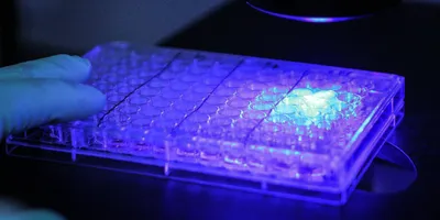
As Murphy describes, “We developed a process that allowed us to test an array of materials—each one slightly different in terms of stiffness or ability to attach to stem cells—on a single slide. It was automated, using a liquid-handling robot, and we could screen hundreds of materials in a month; which we can now do in a week,” adding that in the “olden days,” each experiment would only be able to screen about 10 materials.
A UW-Madison spinoff called Stem Pharm has licensed patents for the materials from the Wisconsin Alumni Research Foundation and is starting to sell the system to pharmaceutical companies and scientific institutes, says Murphy, who is Stem Pharm’s co-founder and chief science officer. He added that “Increasingly, pharmas are externalizing innovation, because internally they don’t have as much capacity to innovate as before. A number of companies have expressed a strong interest in moving away from Matrigel, and our vascular screening product has already been successfully beta-tested at multiple locations.”