Sitting in chemical libraries in labs across the world are compounds waiting to become drugs. When researchers screen hundreds of thousands of these compounds against a specific drug target, a lucky handful might show promise. Some may even overcome every hurdle along the drug discovery pipeline to become an approved drug. Even then, their journey isn’t necessarily complete. They may be combined with other approved or investigational drugs to enhance their efficacy, generating another enormous collection of samples that will get screened all over again.
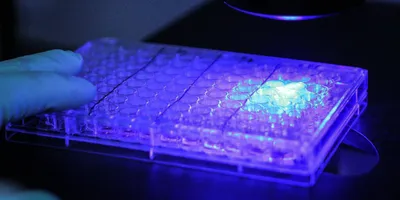
The ability to search a seemingly never-ending sample space in drug screening campaigns depends on miniaturization: decreasing the sample volume used to test a drug’s activity and adapting compound synthesis and sample preparation protocols for a smaller scale. By reducing reagent consumption and cost, miniaturization has made it possible to analyze massive numbers of samples in early-phase drug discovery. The lower material needs of miniaturized systems also enable researchers to screen drugs in cells derived from patients, facilitating translation to the clinic.
As sample volumes approach the nanoliter range in highly miniaturized systems, it’s only natural to wonder if the reliability of the results also downsizes. While shrinking down samples poses both logistical and scientific challenges, researchers have found solutions everywhere from advanced technology to simple laboratory hacks to souped-up statistical analysis. By developing robust assays and respecting their limitations, scientists can ensure that small samples have an enormous impact.
Well, well, well
The key to a robust assay at any scale is rigorous development, optimization, and validation. Before screening drugs in 384- or 1,536-well plates, researchers evaluate an assay’s performance by measuring parameters like signal variation between samples, signal-to-background ratio, and Z-factor (a numerical value that represents its ability to distinguish between positive and negative controls). “We’ll start to miniaturize, and we’ll see how those metrics hold up,” said Douglas Auld, a high-throughput biologist at the Novartis Institutes for BioMedical Research (NIBR).
In a biochemical assay, researchers can tweak the composition of the buffer to enhance the signal window. Holly Yin, a high-throughput screening scientist at the City of Hope National Medical Center, found that adjusting the density of cells seeded into each well and the incubation period can improve these metrics in miniaturized cell-based assays. Researchers can also perform replicate measurements to enhance statistical performance. “An advantage of highly miniaturized assays is that you can potentially look at multiple copies of the library to try to gain increased confidence,” Auld said.
James Inglese, an assay development and screening technology researcher at the National Center for Advancing Translational Sciences, performs replicate measurements at multiple concentrations of each drug compound he tests to generate a dose-response curve during the primary, miniaturized screen. This method, called quantitative high-throughput screening, provides a full efficacy profile for each candidate rather than selecting only those compounds that show activity at a single concentration for further analysis (1). “The reason we can start at the full titration instead of going through all those initial steps is because we miniaturized the system,” Inglese said. “And then we can run that multiple times, because it's just so little material. We can actually get very good reproducibility statistics on each of these measurements.”
Where there’s a will for miniaturization, there’s a way, Inglese said. “If one spends more time thinking about assay design, you can almost guarantee you're going to find a way to come up with a miniaturized version of that assay.”
However, assay development is only one part of the equation. The lower material needs of miniaturized assays have catalyzed strategies to synthesize compounds in smaller quantities, said Cara Brocklehurst, a synthetic chemist at NIBR. Brocklehurst’s team synthesizes compounds designed for a specific drug target using information from previous screens as part of the iterative design-make-test-analyze cycle. To make compounds at a scale appropriate for miniaturized assays, the researchers turn to miniaturization themselves, running 96 discrete reactions in a 96-well plate that each generate about one milligram of a unique product.
While this platform uses an automated workflow, significant effort went into developing the technology before pressing the button. “Doing chemistry on a smaller scale can bring advantages; however, going to the sort of scales that we're talking about actually comes with its own disadvantages because you're having to deal with a lower reaction volume and you're having to purify material on a smaller scale,” Brocklehurst said. For example, the team had to design an entirely new miniaturized purification system and a method to quantify the product by integrating solution component peaks instead of weighing out a solid (2).
Once the compounds are prepared, researchers use advanced liquid handling instrumentation to dispense low volumes of assay reagents into the well plates. “The dispensing technology today is very good,” Inglese said. “It's very expensive equipment, so that’s one of the barriers, but I think in the end, the efficiency makes up for all that over time.”
To perform assays in 1,536-well plates at a volume of 10 microliters, Inglese uses a semi-automated system featuring a dispense head in which reagent is fed through the tops of the tips rather than aspirated in the way of handheld pipettes, enabling uniform, nanoliter-scale dispensing. Yin uses a pipette tip-free instrument that relies on acoustic sound waves to dispense monodisperse droplets. “They dose as little as 2.5 nanoliters, which cannot be pipetted manually at all,” she said.
Small volumes can present challenges even after the samples are prepared. “Evaporation is much more rapid from a small volume than it is from a larger well or a test tube,” Inglese said. To prevent error due to evaporation, Inglese seals his well plates with a heavy lid with small holes for respiration. Yin does not place samples in the wells along the edges of the plate, where evaporation is most likely to occur during longer incubation periods.
Drop it low
While some researchers have explored further miniaturizing screening in even higher density well plates (such as 3,456-well plates containing two microliters of sample), the 1,536-well format is where technology has settled for now. But moving from screening in well plates to microwell chips allows researchers to take sample volumes even lower.
Paul Blainey, a biological engineer at Massachusetts Institute of Technology (MIT), developed a microfluidic droplet-based platform for combinatorial drug discovery in which researchers mix together cells, a drug compound, and a distinct ratio of three fluorescent dyes that provides a unique “barcode” for the compound. After the solutions are emulsified into one-nanoliter droplets, they load two random droplets into each well in a microwell chip and measure the fluorescence output of each droplet to read its barcode and identify the compound it contains. They then apply an electric field to the chip that causes the droplets to merge, and after a reaction period, they image the array to determine the combinatorial effect of the two drugs on the cells.
In a 2018 study in the Proceedings of the National Academy of Sciences, Blainey and colleagues used their platform to screen more than 100,000 combinations of 4,000 investigational and approved drugs and 10 antibiotics for bacterial cell growth inhibition (3). One challenge in microfluidic droplet screening is that drug compounds can escape and transfer between droplets due to the exchange of hydrophobic and hydrophilic molecules at the droplet interface. To minimize cross-contamination between droplets in neighboring wells, the researchers developed a method to rinse and seal the microwell array that limited compound diffusion. They measured each combination of compounds at an average of 13 replicates, which was easy to achieve with their high-throughput system, allowing them to define a threshold at which they could confidently identify hit compounds with few false positives.
This miniaturized system reduces cost by an order of magnitude and compound consumption by two orders of magnitude. Decreasing the amount of compound used is especially critical for combinatorial screening, where the quantity of material required to test each compound against all others can be prohibitive. “It’s not just faster or cheaper; it’s the difference between being able to do it and not being able to do it,” Blainey said .
Researchers at NIBR are developing a similar microdroplet platform called the Mic-Drop to perform assays in droplets less than one nanoliter in volume. (A single drop of water is about 50 microliters, or 50,000 times bigger.) In addition to using Mic-Drop to explore a wide chemical space, the researchers hope this highly miniaturized system will eventually enable screens against scarce, patient-derived cells.
But screening at this scale is only the first step. “When we look at miniaturized screens, especially beyond 1,536-well plates, we don't look at them as a replacement for lower-throughput, more traditional assays,” said Ken Yamada, a chemist at NIBR who is working on the Mic-Drop. “We see this as a complementary approach where we can interrogate a much larger number of compounds or different assays or perhaps the individual cell level that you cannot interrogate in a well plate because you're looking at the average.”
For this reason, miniaturization is not necessary for every application. “There are opportunities afforded, so it's smart to take advantage of those and to select projects that really benefit from those characteristics,” Blainey said.
When miniaturization is appropriate, researchers should identify and operate within the constraints of a specific assay to determine how low they should go, according to Blainey. For example, when his team performs screens against bacteria, the droplet must be large enough to support bacterial growth. The minimum volume for a biochemical assay, on the other hand, might be determined by detection sensitivity. “The constraints are different,” Blainey said, “but the fundamentals don’t change.”
References
- Inglese, J. et al. Quantitative high-throughput screening: A titration-based approach that efficiently identifies biological activities in large chemical libraries. Proc Natl Acad Sci USA 103, 11473-11478 (2006).
- Ginsburg-Moraff, C., et al. Integrated and automated high-throughput purification of libraries on microscale. SLAS Technol (2022). In press.
- Kulesa, A., Kehe, J., Hurtado, J.E., Tawde, P., & Blainey, P.C. Combinatorial drug discovery in nanoliter droplets. Proc Natl Acad Sci USA 115, 6685-6690 (2018).