Research Areas
| 5 min read
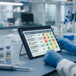
Regulatory Management by Uncountable brings per-ingredient compliance screening into the formulation workflow, flagging issues by market before locking a recipe
| 2 min read
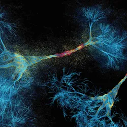
In mice, the new drug led to recovery of motor function and vision by protecting nerve fibers and myelin.
| 2 min read
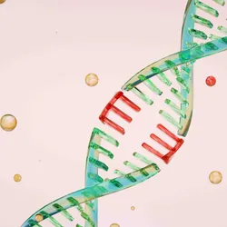
The next-generation tool, called Partita, identified both known and novel genes that contribute to resistance in lymphomas.

| 2 min read
New findings slowed tumor growth in a mouse model of triple-negative breast cancer, offering a new template for RNA drug design.
| 4 min read
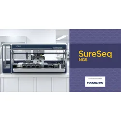
SureSeq NGS workflows on the Hamilton STAR Line cut hands-on library preparation time, giving clinical research labs room to scale throughput
| 4 min read
A win for autoimmune disease, a set back for Alzheimer’s disease, an FDA backing for a multi-cancer blood test, and more led the news this week.

| 3 min read
Autonomous agents flagged a family of phage enzymes with CRISPR-like repeats, though what the system does remains unknown.

| 4 min read
As AI moves into increasingly consequential applications, the next challenge is not what these systems can do, but whether people can trust the answers they provide.

| 4 min read
Proteogenomic analysis of brain tumors identified potential treatment targets and prognostic markers that differed across age, developmental stage, and sex.
| 5 min read
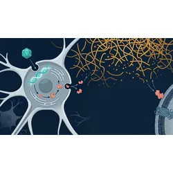
Transpher A data show a 23.5-point cognitive advantage over natural history, supporting the first-ever FDA approval for pediatric patients with MPS IIIA
Popular Topics


Multimedia
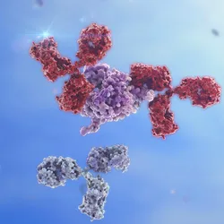
Explore how surface plasmon resonance can reveal binding kinetics, epitope relationships, and receptor interactions to guide antibody lead optimization.


Explore how enzymatic DNA synthesis can support longer, more complex constructs for genome editing, mRNA, and synthetic biology workflows.


Explore how direct cell lysis can reduce sample preparation steps and support efficient gene expression analysis by RT-qPCR.